. 2012 Nov 15;125(Pt 22):5535-45.
doi: 10.1242/jcs.111260. Epub 2012 Sep 12.

Macrophages require Skap2 and Sirpα for integrin-stimulated cytoskeletal rearrangement

Affiliations

Macrophages require Skap2 and Sirpα for integrin-stimulated cytoskeletal rearrangement

Francis J Alenghat et al. J Cell Sci. .

Abstract

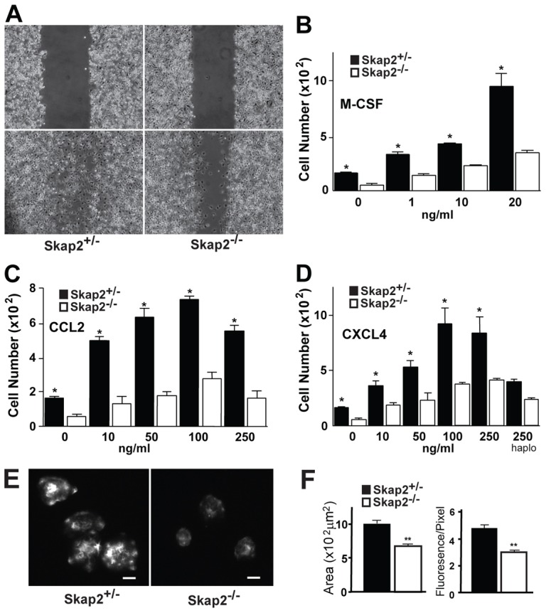
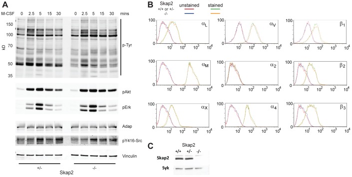
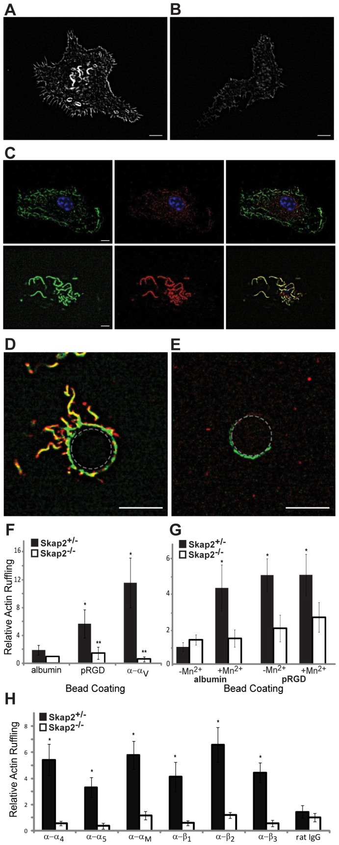
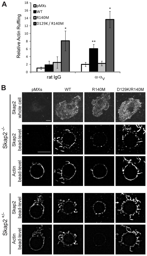
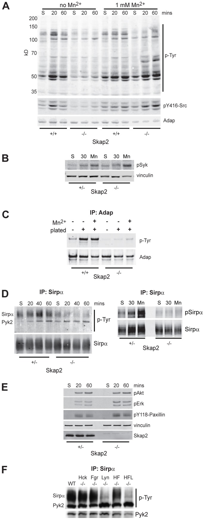
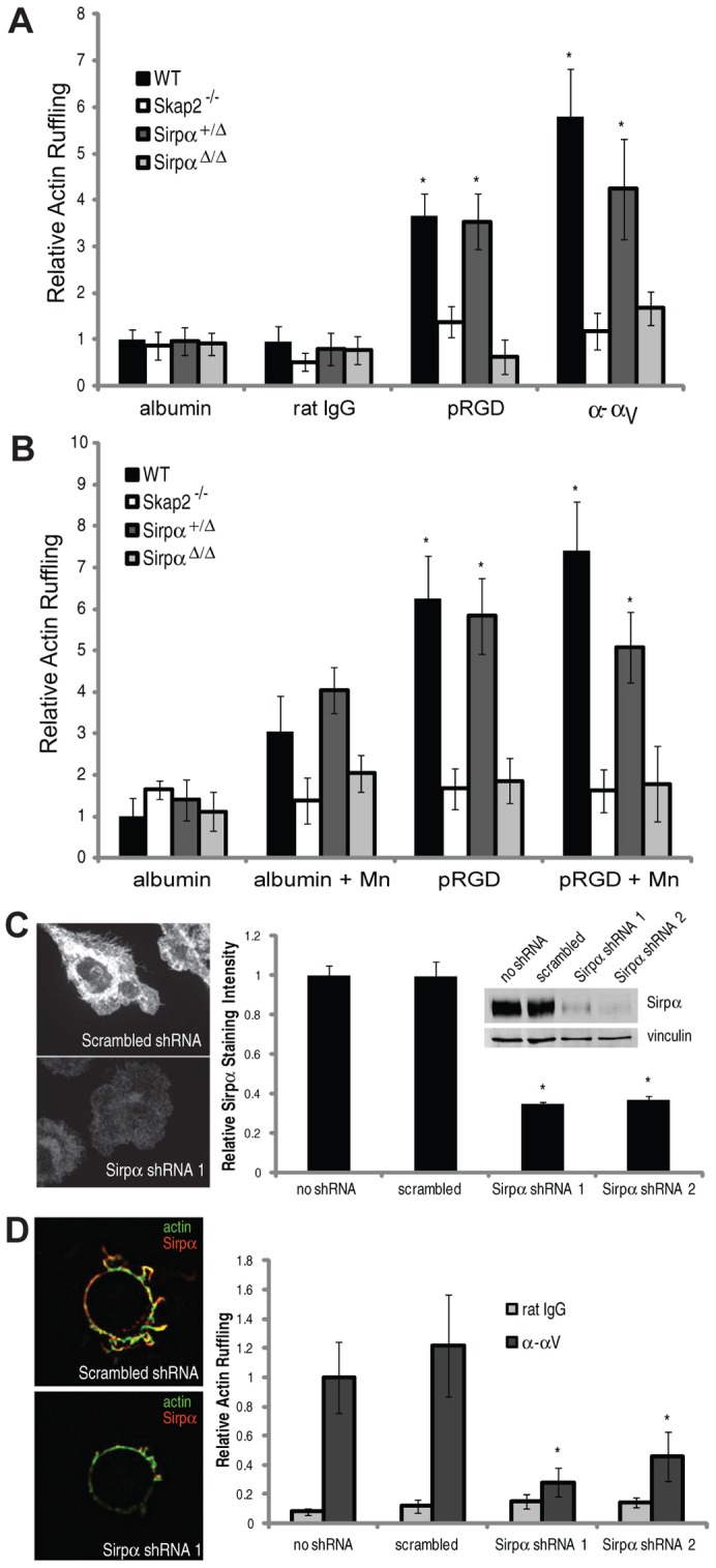
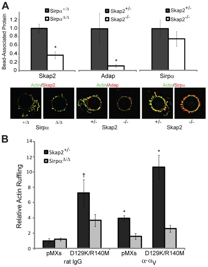
Macrophages migrate to sites of insult during normal inflammatory responses. Integrins guide such migration, but the transmission of signals from integrins into the requisite cytoskeletal changes is poorly understood. We have discovered that the hematopoietic adaptor protein Skap2 is necessary for macrophage migration, chemotaxis, global actin reorganization and local actin reorganization upon integrin engagement. Binding of phosphatidylinositol [3,4,5]-triphosphate to the Skap2 pleckstrin-homology (PH) domain, which relieves its conformational auto-inhibition, is critical for this integrin-driven cytoskeletal response. Skap2 enables integrin-induced tyrosyl phosphorylation of Src-family kinases (SFKs), Adap, and Sirpα, establishing their roles as signaling partners in this process. Furthermore, macrophages lacking functional Sirpα unexpectedly have impaired local integrin-induced responses identical to those of Skap2(-/-) macrophages, and Skap2 requires Sirpα for its recruitment to engaged integrins and for coordinating downstream actin rearrangement. By revealing the positive-regulatory role of Sirpα in a Skap2-mediated mechanism connecting integrin engagement with cytoskeletal rearrangement, these data demonstrate that Sirpα is not exclusively immunoinhibitory, and illuminate previously unexplained observations implicating Skap2 and Sirpα in mouse models of inflammatory disease.


Figures

Fig. 1.
Fig. 1.
Skap2-deficient BMMs exhibit defective migration, spreading, and actin polymerization. (A) Scratches were introduced into confluent cultures of Skap2+/− or Skap2−/− BMMs (upper panels). After 8 hours (lower panels), migration across the scratch wounds was impaired in cells lacking Skap2. (B–D) Chemotaxis of Skap2+/− and Skap2−/− BMMs in response to cytokine M-CSF (B) and chemokines CCL2 (C) and CXCL4 (D) in transwell migration assays. In D, “haplo” denotes haplotaxis conditions where equal concentrations of CXCL4 are in both chambers. For B–D, data are presented as mean ± S.E.M., n = 3, *P<0.01 compared to Skap2−/− under the same condition. (E) Skap2+/− or Skap2−/− BMMs were suspended in DMEM for 3 hours, plated for 30 minutes on glass coverslips, then fixed and stained for F-actin with rhodamine-labeled phalloidin. Scale bar: 10 µm. (F) Quantification of plating-induced spreading area from (E) in µm2 (left) and relative fluorescence intensity per pixel for rhodamine-labeled phalloidin (right) for Skap2+/− and Skap2−/− BMMs. Data presented as mean ± S.E.M., n = 30, **P<0.001 compared to Skap2+/− BMMs.
Fig. 2.
Fig. 2.
Skap2 is not required for M-CSF-induced signaling and does not affect integrin expression in macrophages. (A) Adherent Skap2+/− and Skap2−/− BMMs treated with M-CSF for the indicated times were lysed, electrophoresed, and immunoblotted for phosphotyrosine (p-Tyr) and phosphorylated Akt, Erk2, and Src, with total Adap and Vinculin as loading controls. (B) Flow cytometric analysis was performed on Skap2-replete (WT, except Skap2+/− for αV and β1) or Skap2−/− BMMs using antibodies against the indicated cell surface integrins. (C) Skap2+/+ and Skap2+/− BMMs express equal amounts of Skap2 protein; Syk is a loading control. For all panels, representative results from two independent experiments are shown.
Fig. 3.
Fig. 3.
Skap2 is required for integrin-dependent actin cytoskeletal rearrangement. (A) Confocal micrograph of a typical phalloidin-stained Skap2+/− BMM plated on glass, demonstrating curvilinear actin ruffles. (B) Skap2−/− BMMs typically exhibit less actin ruffle formation. (C) At the plane of the nucleus (top panels, with blue nucleus in focus), a Skap2+/− BMM has minimal cortical actin (green) and cytosolic Skap2 (red), without significant colocalization (yellow, in merge); in contrast, at the apical surface (bottom panels), Skap2 colocalizes strongly with cortical actin ruffles. See also supplementary material Movie 1. (D) Upon binding to a Skap2+/− BMM for 20 minutes, a polystyrene bead (denoted by dotted outline) coated with anti-αV integrin antibodies recruits actin ruffles (green) colocalized with Skap2 (red). See also supplementary material Movie 2. (E) This response is markedly reduced in Skap2−/− BMMs. Scale bar: 10 µm. (F) Quantified actin ruffling responses, measured as average pixel intensity in an 8-µm annulus surrounding beads coated with integrin-engaging or control proteins, are shown for BMMs of both genotypes. Data are normalized against the albumin/Skap2−/− condition as Relative Actin Ruffling and presented as mean ± S.E.M., n = 10 per condition, *P<0.05 compared to albumin-coated beads on the same cells, **P<0.01 compared to same beads on Skap2+/− BMMs. (G) Quantified actin ruffling responses, induced by binding to polystyrene beads coated with either albumin or polyRGD, is shown for Skap2+/− and Skap2−/− BMMs with or without treatment with 1 mM Mn2+. Data are normalized against the Mn2+/albumin/Skap2+/− condition and presented as mean ± S.E.M., n = 10 per condition, *P<0.05 compared to albumin-coated beads, no Mn2+, on the same cells. (H) Quantified actin ruffling responses to subtype-specific integrin-engaging beads are shown for BMMs of both genotypes. Data are normalized against the rat IgG/Skap2−/− condition and presented as mean ± S.E.M., n = 10 per condition, *P<0.01 compared to the same beads on Skap2−/− BMMs.
Fig. 4.
Fig. 4.
Integrin-induced cytoskeletal reorganization requires a functional Skap2 PH domain. (A) Actin ruffling induced by beads coated with rat IgG (control) or anti-αV bound to Skap2−/− BMMs infected with empty vector (pMXs), WT Skap2 (WT), or Skap2 mutants with an R140M substitution or with combined D129K and R140M substitutions. Data are presented as mean ± S.E.M., n = 10 per condition, *P<0.01 compared to pMXs, **P<0.05 compared to pMXs. (B) Skap2−/− and Skap2+/− BMMs infected with empty vector (pMXs), WT Skap2 (WT), or Skap2 mutants with an R140M substitution or with combined D129K and R140M substitutions were bound to polystyrene beads coated with antibodies directed against αV integrin, and stained for actin (phalloidin) and for Skap2. n = 10 per condition. Scale bar: 10 µm.
Fig. 5.
Fig. 5.
Skap2 is required for specific integrin-induced signaling events. (A) Skap2+/+ and Skap2−/− BMMs in suspension (S) or plated on fibrinogen for 20 or 60 minutes, either in the absence (left six lanes) or presence (right six lanes) of 1 mM Mn2+, were lysed, electrophoresed and immunoblotted for phosphotyrosine (p-Tyr; upper panel), Src pY416 (middle panel), and Adap as a loading control (lower panel). (B) Skap2+/− and Skap2−/− BMMs were kept in suspension or plated on fibrinogen for 30 minutes in the absence or presence of Mn2+, lysed, and immunoblotted for pSyk and vinculin as a loading control. Baseline quantified pSyk/vinculin is ∼2-fold higher in Skap2−/− cells. Plating increases levels ∼2-fold and Mn2+ increases levels ∼5-fold in both genotypes. (C) Skap2+/+ and Skap2−/− BMMs were kept in suspension or plated on fibrinogen for 30 minutes in the absence or presence of Mn2+, lysed, immunoprecipitated for Adap, and immunoblotted for phosphotyrosine (pTyr; upper panel) and Adap (lower panel). Baseline quantified pTyr/Adap is ∼5-fold higher in Skap2+/− cells. Plating increases levels ∼5-fold in Skap2+/− and ∼1.2-fold in Skap2−/− cells. Mn2+ increases levels ∼6-fold in Skap2+/− cells and 3-fold in Skap2−/− cells. (D) Sirpα was immunoprecipitated from lysates of BMMs in suspension (S) or after plating for the indicated times on fibrinogen, and phosphotyrosine (p-Tyr) was analyzed by immunoblotting (left panels), with prominent bands corresponding to Sirpα and Pyk2. Similar lysates of suspended, plated, or 1 mM Mn2+-treated BMMs (right panels) were probed for Sirpα phosphorylation. (E) Akt, Erk, and Paxillin phosphorylation were probed in Skap2+/− and Skap2−/− BMMs in suspension (S) or after plating for the indicated times on fibrinogen. (F) BMMs from Hck−/−, Fgr−/−, and Lyn−/−, as well as Hck/Fgr double knockout (HF−/−) and Hck/Fgr/Lyn triple knockout (HFL−/−) mice, were lysed after plating on fibrinogen, subjected to immunoprecipitation for Sirpα, and immunoblotted for p-Tyr and Pyk2. All panels are representative of at least three independent experiments.
Fig. 6.
Fig. 6.
Integrin-induced cytoskeletal reorganization is also Sirpα-dependent. (A) Actin ruffling induced by beads coated with albumin, rat polyclonal IgG, pRGD, or anti-αV bound to WT, Skap2−/−, Sirpα+/Δ, and SirpαΔ/Δ BMMs. (B) Actin ruffling induced by beads coated with albumin or pRGD with or without 1 mM Mn2+. For A and B, data are presented as mean ± S.E.M., n = 10 per condition, *P<0.01 compared to either homozygous mutant cell under same conditions. (C) Immunofluorescence staining for Sirpα in representative WT BMMs treated with scrambled versus Sirpα shRNA, along with quantified relative staining intensity in untreated cells and cells treated with scrambled shRNA versus the two different Sirpα shRNA. Inset shows western analysis of cell lysates of the same cell populations, blotted for Sirpα. (D) Immunofluorescence staining for actin and Sirpα in response to αV-directed beads on WT BMMs treated with indicated shRNA, along with quantified actin ruffling induced by these beads. Data are presented as mean ± S.E.M., n = 10 per condition, *P<0.05 compared to scrambled shRNA and untreated conditions.
Fig. 7.
Fig. 7.
Sirpα and Skap2 cooperate at sites of integrin engagement to drive cytoskeletal rearrangement. (A) Quantified Skap2 recruitment to α–αV beads bound to Sirpα+/Δ and SirpαΔ/Δ BMMs, and Adap and Sirpα recruitment to α–αV beads bound to Skap2+/− and Skap2−/− BMMs. Data are normalized to the respective heterozygous condition and presented as mean ± S.E.M., n = 10–20 per condition, * P<0.05. Representative beads, co-stained for actin, are shown. (B) Actin ruffling induced by beads coated with rat polyclonal IgG or anti-αV, bound to Skap2+/− and SirpαΔ/Δ BMMs infected with empty vector (pMXs) or D129K/R140M Skap2. Data are presented as mean ± S.E.M., n = 10–15 per condition; † P = 0.1 compared to SirpαΔ/Δ and P<0.01 compared to pMXs under the same conditions; * P<0.01 compared to SirpαΔ/Δ under the same conditions.
Fig. 8.
Fig. 8.
Skap2 and Sirpα in integrin signaling. A Sirpα/Skap2/Adap signaling module drives actin cytoskeleton reorganization downstream of integrin engagement in macrophages.

Similar articles

Cited by

References

    1. Adams S., van der Laan L. J., Vernon–Wilson E., Renardel de Lavalette C., Döpp E. A., Dijkstra C. D., Simmons D. L., van den Berg T. K. (1998). Signal-regulatory protein is selectively expressed by myeloid and neuronal cells. J. Immunol. 161, 1853–1859 - PubMed
    1. Alenghat F. J., Tytell J. D., Thodeti C. K., Derrien A., Ingber D. E. (2009). Mechanical control of cAMP signaling through integrins is mediated by the heterotrimeric Galphas protein. J. Cell. Biochem. 106, 529–538 10.1002/jcb.22001 - DOI - PMC - PubMed
    1. Barclay A. N. (2009). Signal regulatory protein alpha (SIRPalpha)/CD47 interaction and function. Curr. Opin. Immunol. 21, 47–52 10.1016/j.coi.2009.01.008 - DOI - PMC - PubMed
    1. Berton G., Lowell C. A. (1999). Integrin signaling in neutrophils and macrophages. Cell. Signal. 11, 621–635 10.1016/S0898-6568(99)00003-0 - DOI - PubMed
    1. Berzat A., Hall A. (2010). Cellular responses to extracellular guidance cues. EMBO J. 29, 2734–2745 10.1038/emboj.2010.170 - DOI - PMC - PubMed

Publication types

MeSH terms

Substances