. 2012 Dec 1;125(Pt 23):5667-76.
doi: 10.1242/jcs.107490. Epub 2012 Sep 12.

Transcriptional regulation of Profilin during wound closure in Drosophila larvae

Affiliations

Transcriptional regulation of Profilin during wound closure in Drosophila larvae

Amanda R Brock et al. J Cell Sci. .

Abstract

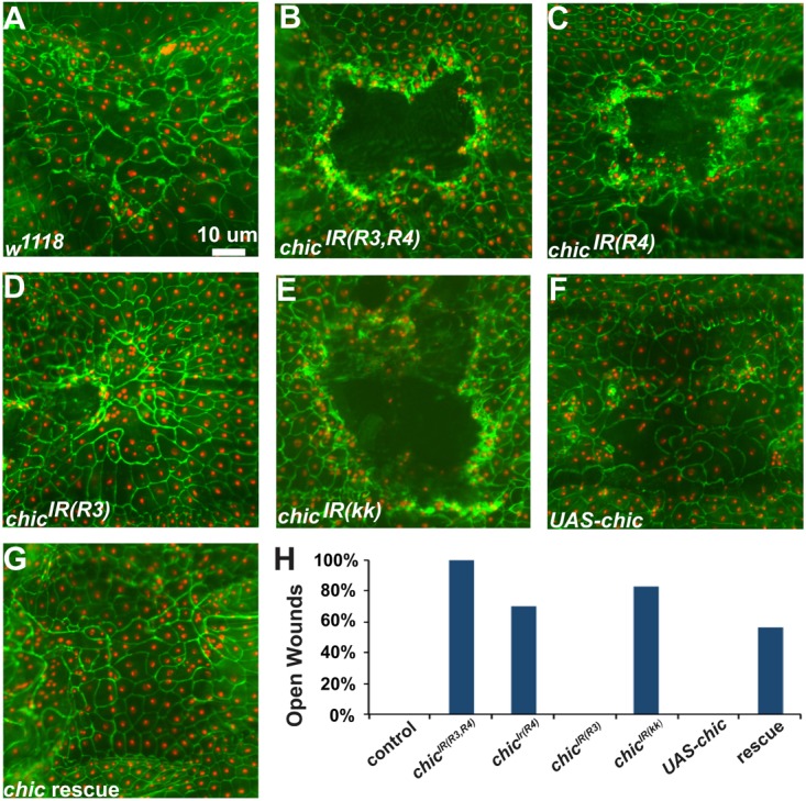
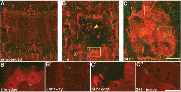
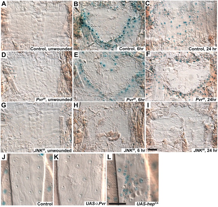
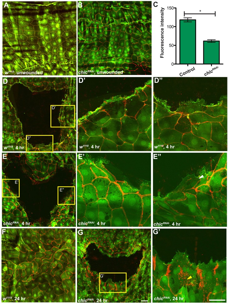
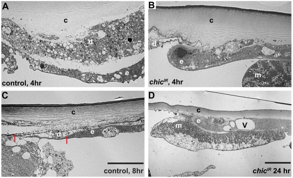
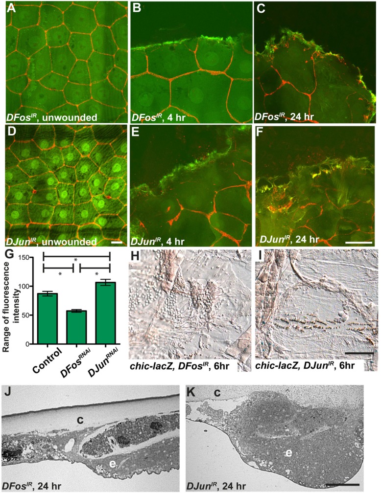
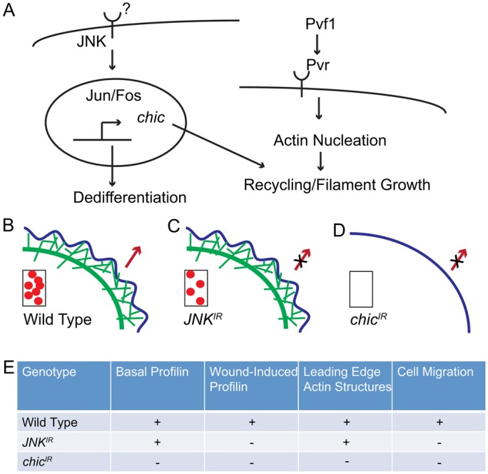
Injury is an inevitable part of life, making wound healing essential for survival. In postembryonic skin, wound closure requires that epidermal cells recognize the presence of a gap and change their behavior to migrate across it. In Drosophila larvae, wound closure requires two signaling pathways [the Jun N-terminal kinase (JNK) pathway and the Pvr receptor tyrosine kinase signaling pathway] and regulation of the actin cytoskeleton. In this and other systems, it remains unclear how the signaling pathways that initiate wound closure connect to the actin regulators that help execute wound-induced cell migrations. Here, we show that chickadee, which encodes the Drosophila Profilin, a protein important for actin filament recycling and cell migration during development, is required for the physiological process of larval epidermal wound closure. After injury, chickadee is transcriptionally upregulated in cells proximal to the wound. We found that JNK, but not Pvr, mediates the increase in chic transcription through the Jun and Fos transcription factors. Finally, we show that chic-deficient larvae fail to form a robust actin cable along the wound edge and also fail to form normal filopodial and lamellipodial extensions into the wound gap. Our results thus connect a factor that regulates actin monomer recycling to the JNK signaling pathway during wound closure. They also reveal a physiological function for an important developmental regulator of actin and begin to tease out the logic of how the wound repair response is organized.


Figures

Fig. 1.
Fig. 1.
chicRNAi results in a failure of wound closure. (A-G) Dissected larval epidermal wholemounts stained for anti-Fasciclin III (green). Genotype is w; e22c-gal4, UAS-dsred2nuc (to label epidermal nuclei, red) plus the indicated mutations or UAS transgenes. All panels show wounded larvae 24 hours after wounding. (A) w1118, control. Note the presence of large, atypically shaped cells, some of which are multinucleate, at the closed wound (compare with unwounded epidermis in Fig. 2A). (B) UAS-chicIR(R3,R4). Note the open wound gap. (C) UAS-chicIR(R4). Note the open wound gap. (D) UAS-chicIR(R3). Note the closed wound. (E) UAS-chicIR(kk). Note the open wound gap. (F) UAS-chickadee. Overexpression of chickadee does not interfere with wound closure. (G) UAS-chicIR(R3,R4), UAS-chickadee. A closed wound is shown. (H) Percentage of larvae with an open wounds versus genotype. Rescue refers to UAS-chicIR(R3R4), UAS-chickadee. n≥30. Scale bar: 10 µm.
Fig. 2.
Fig. 2.
Profilin relocalizes and its levels increase following wounding. (A–C″) Dissected larval epidermal wholemounts of genotype: w;dmef2-gal4, FasIII-GFP/+;UAS-chicIR(R3,R4)/+. In these larvae, the muscle Gal4 driver dmef2-Gal4 drives expression of chicIR(R3,R4) in order to knock down muscle Profilin expression that would obscure a clear view of the epidermal Profilin. Epidermal membranes are labeled with FasIII-GFP (green); Profilin is labeled by α-chic antibody (red). (A) Unwounded larval epidermis. Note low levels of perinuclear Profilin staining. (B) Larvae at 6 hours after wounding. Note that in cells adjacent to the wound edge, Profilin levels have significantly increased (B′) compared with cells located several cell rows away from the wound (B″). Note that the Profilin antibody also labels blood cells (yellow arrowhead) in the middle of the wound. (C) Larvae at 24 hours after wounding. Profilin is still high in cells that have migrated to close the wound gap, as indicated by the sharp boundary of the former wound area (C′). Levels are still high within the former wound (C″). Note: Due to the extreme disparity in fluorescence levels between control and wounded samples red-channel levels were adjusted to different optimal levels for these samples. Scale bars: 100 µm (A,B,C); 50 µm (B′,B″,C′,C″).
Fig. 3.
Fig. 3.
chic transcription is regulated by the JNK but not by the Pvr signaling pathway. (A–I) Dissected epidermal wholemounts of larvae heterozygous for w;e22c-Gal4, chic01320 and the indicated mutants or transgenes. All are stained with X-Gal (see Materials and Methods) to highlight β-Galactosidase activity in lacZ-expressing nuclei (blue). (A–C) w1118, (D–F) UAS-PvrRNAi or (G–I) UAS-JNKRNAi. (A,D,G) Unwounded, (B,E,H) 6 hours after wounding and (C,F,I) 24 hours after wounding. Note the increase in lacZ expression in wild type (B) following wounding. This increase is absent in cells lacking JNK expression (H). (JL) Dissected epidermal wholemounts of larvae of genotype: w;tubgal80ts/chic01320; pnr-Gal4, UAS-GFP (J) plus UAS-λPvr (K), or UAS-hepCA (L). Note the induction of lacZ in larvae with constitutively activated JNK expression (L). Scale bars: 100 µm (A–I); 50 µm (J–L).
Fig. 4.
Fig. 4.
Actin localizes to the wound edge and forms processes in wild type but not in larvae lacking Profilin expression. (A,B,D-G) Dissected epidermal wholemounts of larvae heterozygous for w;e22c-gal4, UAS-lifeact-GFP6.0 and the indicated mutations or transgenes. Epidermal membranes are labeled with FasIII (red). (A,D,F) w1118 (control). (B,E–E″,G,G′) chicIR(R3,R4). (A,B) Unwounded, (D,E′′) 4 hours after wounding and (F,G′) 24 hours after wounding. Yellow boxes indicate the areas shown at higher magnification in D′,D″, E′, E″ and G′. Note the discontinuous actin cable in D and the presence of actin-based extensions in D′ and D″. Note that the wound is closed after 24 hours (F). Note the lack of actin cable in E, E′, G and G′. The white arrowhead in E″ indicates a small actin-based process. The yellow arrowhead in G′ represents presumptive cytoplasmic vesicles. Note that wounds are open at 24 hours (G,G′). (C) Graph shows the range of the fluorescence intensity between the leading edge and the interior of the cell in both control and chicIR(R3,R4) at 4 hours following wounding. Cable intensity is significantly different between groups (*P≤0.01, Student's t-test). Error bars represent the s.e.m. For the control group n = 96 measurements; for chicIR, n = 64. Scale bars: 50 µm.
Fig. 5.
Fig. 5.
chic is required for cells to extend processes into the wound gap. (A–D) TEM images of transverse sections of wounded and dissected L3 larvae of genotypes: w; UAS-nlacZ/+; UAS-chicIR(R3,R4) (A); w; e22c-Gal4/UAS-nlacZ; UAS-chicIR(R3,R4) (B,D); and w;pxn-Gal4, UAS-nlacZ/+ (C). In all images, the wound gap is to the left and the cells are migrating in that direction. (A,B) Larvae at 4 hours after wounding. Note that the control extends a long thin process over the necrotic cellular debris (A), whereas cells lacking Profilin form a rounded and blunt wound edge (B). (C) Larvae at 8 hours after wounding. The cell extends a normal process that is 14.4 µm long (between the red marks) and 0.4 µm thick. (D) Larvae at 24 hours after wounding. The wound has still not closed and the rounded cell has not appreciably extended into the wound gap. c, cuticle; m, muscle; e, epidermal cell; d, cell debris; V, large cytoplasmic vesicle. Scale bars: 10 µm.
Fig. 6.
Fig. 6.
DFos and DJun are required for chic transcription. (A–F) Dissected larval epidermal wholemounts of genotype w;e22c-Gal4, UAS-lifeactGFP (to label epidermal actin in green) plus UAS-DFosIR (A–C) or UAS-DJunIR (D–F) and stained for Fasciclin III (red). (A,D) Unwounded, (B,E) and 4 hours after wounding. Note that at 4 hours, the DFosIR-expressing larva (B) has a dim actin cable, but does not appear to have processes, whereas DJunIR-expressing larvae (E) resemble wild type. (C,F) Larvae at 24 hours after wounding. (G) Quantification of ranges of fluorescence intensity of the actin cable at 4 hours after wounding in the indicated genotypes. Cable intensity is significantly different between each group (*P≤0.01, Student's t-test). Error bars represent s.e.m. For control, n = 145, for DFosIR, n = 146, and for DJunIR, n = 100. (H,I) Dissected larval epidermal wholemounts of genotype w;e22c-Gal4, chic-lacZ plus UAS-DFosIR (H) or UAS-DJunIR (I) at 6 hours after wounding. Note the lack of lacZ induction. (J,K) TEM of transverse sections of wounded and dissected larvae of genotype w;A58-Gal4, UAS-nlacZ/UAS-DFosIR (J) or w; A58-Gal4, UAS-nlacZ/UAS-DJunIR (K) at 24 hours after wounding. e, epidermal cells; c, cuticle. Scale bars: 50 µm (A–F); 100 µm (H,I); 10 µm (J,K).
Fig. 7.
Fig. 7.
Model of JNK and Pvr signaling pathways during wound closure. (A) JNK signaling is shown on the left, with an as-yet unidentified extracellular ligand activating the intracellular JNK, which can in turn activate Jun and Fos. These transcription factors play a role in tissue dedifferentiation required for wound closure. Pvr signaling (shown on the right) activates actin nucleation. Here, we demonstrate that the JNK pathway converges on regulation of actin dynamics by activating transcription of the actin recycling factor chickadee, which is also required for wound closure. (B–D) Visual representation of the actin cytoskeleton (green) at the leading edge of the wound. (B) Wild type; (C) JNKIR; (D) chicIR. Blue line, plasma membrane. Red arrow, direction of migration. In all three cells, the box in the lower left indicates Profilin (red dots) levels. (E) Table summarizing the Profilin expression and actin-based characteristics of the above genotypes.

Similar articles

Cited by

References

    1. Adachi–Yamada T., Fujimura–Kamada K., Nishida Y., Matsumoto K. (1999). Distortion of proximodistal information causes JNK-dependent apoptosis in Drosophila wing. Nature 400, 166–169 10.1038/22112 - DOI - PubMed
    1. Babcock D. T., Brock A. R., Fish G. S., Wang Y., Perrin L., Krasnow M. A., Galko M. J. (2008). Circulating blood cells function as a surveillance system for damaged tissue in Drosophila larvae. Proc. Natl. Acad. Sci. USA 105, 10017–10022 10.1073/pnas.0709951105 - DOI - PMC - PubMed
    1. Berger S., Schäfer G., Kesper D. A., Holz A., Eriksson T., Palmer R. H., Beck L., Klämbt C., Renkawitz–Pohl R., Onel S. F. (2008). WASP and SCAR have distinct roles in activating the Arp2/3 complex during myoblast fusion. J. Cell Sci. 121, 1303–1313 10.1242/jcs.022269 - DOI - PubMed
    1. Boutros M., Paricio N., Strutt D. I., Mlodzik M. (1998). Dishevelled activates JNK and discriminates between JNK pathways in planar polarity and wingless signaling. Cell 94, 109–118 10.1016/S0092-8674(00)81226-X - DOI - PubMed
    1. Brand A. H., Perrimon N. (1993). Targeted gene expression as a means of altering cell fates and generating dominant phenotypes. Development 118, 401–415 - PubMed

Publication types