. 2012 Oct;7(10):1808-17.
doi: 10.1038/nprot.2012.105. Epub 2012 Sep 13.

Dual fluorescence detection of protein and RNA in Drosophila tissues

Affiliations

Dual fluorescence detection of protein and RNA in Drosophila tissues

Hila Toledano et al. Nat Protoc. 2012 Oct.

Abstract

Detection of RNAs by in situ hybridization (ISH) is a well-established technique that permits the study of specific RNA expression patterns in tissues; however, not all tissues are equally amenable to staining using the same procedure. Here we describe a protocol that combines whole-mount immunofluorescence (IF) and fluorescence in situ hybridization (FISH) for the simultaneous detection of specific RNA transcripts and proteins, greatly enhancing the spatial resolution of RNA expression in complex, intact fly tissues. To date, we have successfully used this protocol in adult testis, larval male gonads, adult intestine and Malpighian tubules. IF is conducted in RNase-free solutions, prior to the harsh conditions of FISH, in order to preserve protein antigenicity within dissected tissues. Separate protocols are described for mRNA and miRNA detection, which are based on robust digoxigenin (DIG) RNA and locked nucleic acid (LNA) probes, respectively. The combined IF-FISH procedure can be completed in 2 d for miRNA detection and 4 d for mRNA detection. Although optimized for Drosophila, this IF-FISH protocol should be adaptable to a wide variety of organisms, tissues, antibodies and probes, thus providing a reliable and simple means to compare RNA and protein abundance and localization.


Conflict of interest statement

COMPETING FINANCIAL INTERESTS The authors declare no competing financial interests.

Figures

Figure 1
Figure 1
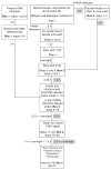
Summary of steps involved in dual fluorescence detection of protein and mRNA/miRNA and the approximate time needed. Dashed boxes indicate that probe preparation is recommended at least 1 d before beginning the protocol. Gray boxes indicate the timing for miRNA detection.
Figure 2
Figure 2
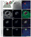
Dual labeling of the stem cell niche in adult and larval Drosophila testes. (a) Schematic showing the apical tip of the Drosophila testis. Germline stem cells (GSCs) and cyst stem cells (CySCs) surround and are in contact with hub cells, which express upd. (b,c) Hybridization of an adult testis (from a 1-d-old male fly) with an RNA probe for the self-renewal factor upd. (b) Detection of DIG-labeled upd riboprobe with the AP–based procedure. The AP substrate precipitates diffusely at sites of AP enzyme activity as shown by a homogenous stain (black) of hub cells. (c) Detection of DIG-upd riboprobe (red) with tyramide signal amplification (TSA) procedure. The peroxidase at the HRP-linked probe reacts rapidly with Tyramine, which results in a much increased spatial resolution of upd transcript detection. DAPI (blue) marks the nuclei of the cells at the apical tip of the testis. Note that the testis appears larger in c because of reduced tissue dehydration during processing compared with b. (df) Dual labeling with antibodies to the germ cell–specific marker Vasa (d) and FISH for upd using a biotin-labeled riboprobe (e); a merged image of Vasa (green) and upd (red) is shown in panel f. (g,h) Dual labeling with antibodies to the hub cell marker Fas3 (g) and FISH for DIG-upd (h). (i) Merged panel showing Fas3 (green), DIG-upd (red) and DAPI (blue). (j,k) Dual labeling for Fas3 (j) and FISH for DIG-upd (k) of third-instar larval (L3) male gonads. (l) Merged panel showing Fas3 (green), DIG-upd (red) and DAPI (blue); arrows point to background signal from the fat body surrounding the larval gonad. Genotype of all imaged flies is OregonR. All panels are single sections. Scale bars, 20 μm.
Figure 3
Figure 3
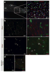
Dual labeling of the Drosophila midgut and Malpighian tubules. (a) Shown are (left) an adult Drosophila gut viewed with a ×10 objective and stained with DAPI showing the posterior midgut (PM), Malpighian tubules (MT), pylorus (Pyl) and hindgut (H), and (right) a higher-magnification view of the posterior midgut showing an intestinal stem cell (ISC)/enteroblast (EB) nest (bracket), a polyploid enterocyte (*) and a Prospero + enteroendocrine cell (arrowhead). (b) Dual labeling with FISH for upd (red) and IF for GFP expression in ISCs/EBs (green) of an adult midgut from a fly of genotype esgGAL4, UAS-gfp. Note upd expression in rows of circular muscle. (c,d) Dual labeling with FISH for esg (red) and IF for GFP (green) of an adult midgut (c) or Malpighian tubules (d). Note colocalization of esg mRNA and the GFP reporter in both tissues (b,c). Scale bars, 10 μm.
Figure 4
Figure 4
FISH in combination with multiple antibodies during IF. (ad) Apical tip of the testis (genotype: esgGAL4, UAS-gfp) showing upd mRNA expression in the hub (a) coimmunolabeled for the cell-cell adhesion marker Armadillo (Arm; b) and for GFP (c); merged image showing upd (red), Arm (blue) and GFP (green) is shown in d. Note upd and Arm colocalization in the hub. (eh) Apical tip of testis showing esg mRNA (e) coimmunolabeled with Arm (f) and GFP (g); merged image showing esg (red), Arm (blue) and GFP (green) is shown in h. Note the difference in esg mRNA expression and expression of GFP driven from the esgGAL4 reporter line. Scale bars, 20 μm.
Figure 5
Figure 5
Dual labeling of let-7 miRNA and Fas3 protein in the testis. An LNA-let-7-DIG-labeled probe was used to detect let-7 miRNA expression in adult testes from 1-d-old male flies (genotype: updGAL4; UAS-let7701.12.9). (ac) Fas3 (a), let-7 (b) and a merged panel, showing Fas3 (green), let-7 (red) and DAPI (blue) (c). Scale bar, 10 μm.

Similar articles

Cited by

References

    1. Speel EJ, Hopman AH, Komminoth P. Amplification methods to increase the sensitivity of in situ hybridization: play card(s) J Histochem Cytochem. 1999;47:281–288. - PubMed
    1. Speel EJ, Saremaslani P, Roth J, Hopman AH, Komminoth P. Improved mRNA in situ hybridization on formaldehyde-fixed and paraffin-embedded tissue using signal amplification with different haptenized tyramides. Histochem Cell Biol. 1998;110:571–577. - PubMed
    1. Baldino F, Chesselet MF, Lewis ME., Jr High-resolution in situ hybridization histochemistry. Methods Enzymol. 1989;168:761–777. - PubMed
    1. Springer JE, Robbins E, Gwag BJ, Lewis ME, Baldino F., Jr Non-radioactive detection of nerve growth factor receptor (NGFR) mRNA in rat brain using in situ hybridization histochemistry. J Histochem Cytochem. 1991;39:231–234. - PubMed
    1. Bobrow MN, Litt GJ, Shaughnessy KJ, Mayer PC, Conlon J. The use of catalyzed reporter deposition as a means of signal amplification in a variety of formats. J Immunol Methods. 1992;150:145–149. - PubMed

Publication types

MeSH terms

LinkOut - more resources