. 2012 Dec 7;12(23):4976-85.
doi: 10.1039/c2lc40338b.

Formation and optogenetic control of engineered 3D skeletal muscle bioactuators

Affiliations

Formation and optogenetic control of engineered 3D skeletal muscle bioactuators

Mahmut Selman Sakar et al. Lab Chip. .

Abstract

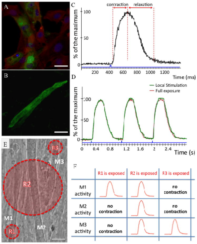
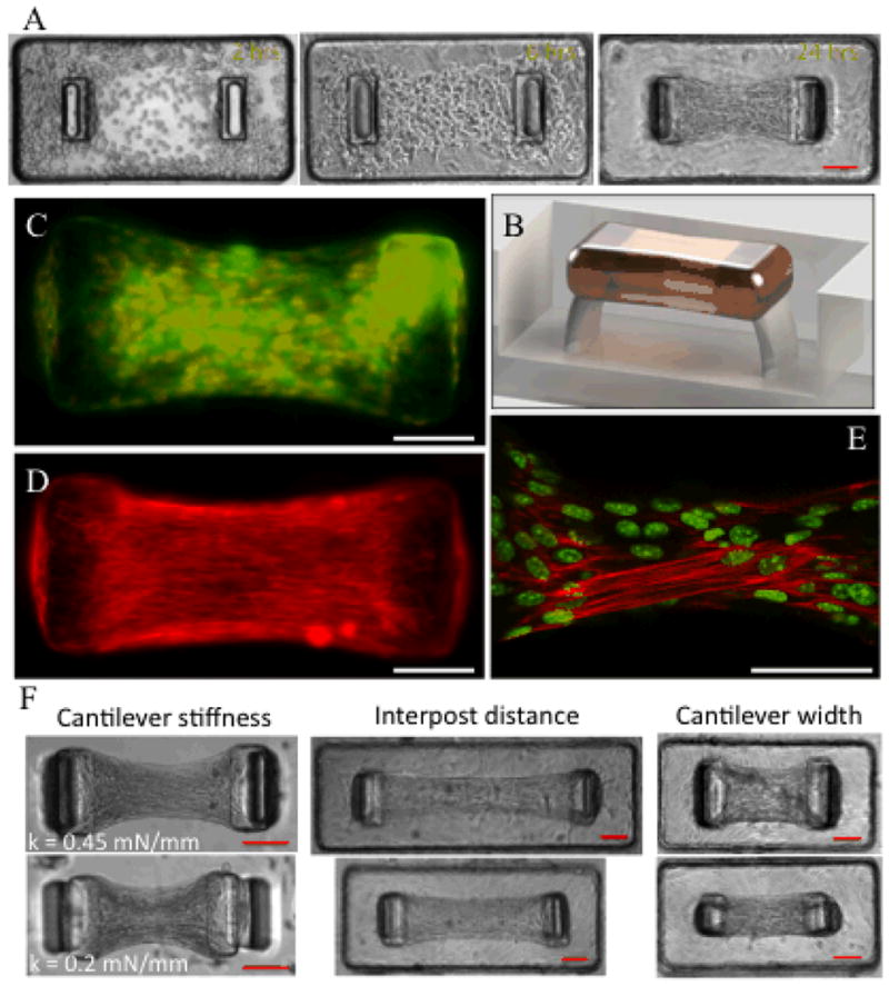
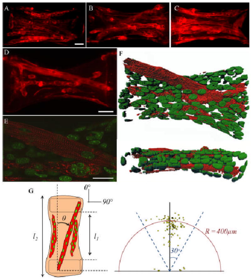
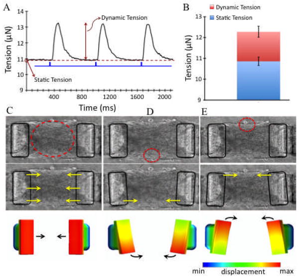
Densely arrayed skeletal myotubes are activated individually and as a group using precise optical stimulation with high spatiotemporal resolution. Skeletal muscle myoblasts are genetically encoded to express a light-activated cation channel, Channelrhodopsin-2, which allows for spatiotemporal coordination of a multitude of skeletal myotubes that contract in response to pulsed blue light. Furthermore, ensembles of mature, functional 3D muscle microtissues have been formed from the optogenetically encoded myoblasts using a high-throughput device. The device, called "skeletal muscle on a chip", not only provides the myoblasts with controlled stress and constraints necessary for muscle alignment, fusion and maturation, but also facilitates the measurement of forces and characterization of the muscle tissue. We measured the specific static and dynamic stresses generated by the microtissues and characterized the morphology and alignment of the myotubes within the constructs. The device allows testing of the effect of a wide range of parameters (cell source, matrix composition, microtissue geometry, auxotonic load, growth factors and exercise) on the maturation, structure and function of the engineered muscle tissues in a combinatorial manner. Our studies integrate tools from optogenetics and microelectromechanical systems (MEMS) technology with skeletal muscle tissue engineering to open up opportunities to generate soft robots actuated by a multitude of spatiotemporally coordinated 3D skeletal muscle microtissues.


Figures

Figure 1
Figure 1
Generation and light-induced stimulation of ChR2-expressing skeletal muscle cells in vitro. (A) Fluorescence image of the membrane-bound ChR2-GFP signal (green) overlaid with F-actin immunostaining (red) in C2C12 myoblasts. Nuclei are shown in blue. (B) Multi-nucleated myotubes expressing the GFP signal (green). (C) Typical contraction pattern of a ChR2-GFP expressing myotube upon stimulation with blue light pulses (10 mW mm−2) for durations indicated by blue bars. A representative example of 20 experiments is shown. (D) Representative repetitive contraction pattern evoked by local and full exposure of a ChR2-GFP–expressing myotube with blue light pulses (10 mW mm−2) for durations indicated by blue bars. (E) Selective activation of myotubes with local stimulation. Myotubes are denoted by M1, M2 and M3 and regions of exposure are labeled with R1, R2, and R3. (F) Blue light pulses confined to a region stimulate only the myotubes residing inside that specific region. (Scale bars: A, 10 μm; B and E, 20 μm).
Figure 2
Figure 2
Generation of skeletal muscle microtissues (SMTs) tethered to elastic force sensors. (A) Representative images depicting the time course of a contracting SMT. (B) Cross section view of the CAD modeling of a single SMT. (C) Representative immunofluorescence overlay of membrane-bound GFP signal (green) and nuclei staining (red) within microtissues showing uniform cell distribution. (D) Representative F-actin imaging (red) revealing the alignment of skeletal muscle myoblasts in the direction of mechanical stress gradients within the microtissues after 3 days of culture. (E) Remodeling of actin (red) within multi-nucleated myotubes. Nuclei are shown in green. (F) Effect of PDMS cantilever stiffness and device geometry on the skeletal muscle microtissue dimensions. (Scale bars: 100 μm).
Figure 3
Figure 3
Distribution and differentiation of skeletal muscle myotubes in engineered microtissues. (A–C) Representative α-actinin immunostaining images showing the distribution and alignment of striated myotubes after 3 weeks of culture. (D) Representative α-actinin immunostaining image shows sarcomere formation and (E) a representative confocal section from the same construct shows that aligned multinucleated myotubes exhibit ubiquitous cross-striations. (F) 3D reconstruction of confocal slices for the construct shown in (D). The upper panel shows the top and the lower panel shows the side view of the same microtissue. Nuclei (green) are elongated in the direction of stress gradients. (G) Characterization of cell alignment and myotube length in the microtissues. The location of each point in the scatter plot shows the length and orientation of a myotube. Data are collected from 20 SMTs having a total of 150 myotubes. (Scale bars: A and D, 50 μm; E, 25 μm).
Figure 4
Figure 4
Functional properties of SMTs and multi degrees of freedom actuation with local stimulation (A) Representative recording of the static and dynamic tension of an SMT on day 15. The microtissue is stimulated with a brief blue light pulse series (indicated by blue bars). (B) Average static and dynamic tension for SMTs at day 15. Data are the average of 50 SMTs ± SEM. (C–E) Multi DOF actuation. Caps are outlined with black rectangles to emphasize motion. Heat maps depicting degree of displacement.

Similar articles

Cited by

References

    1. McMahon TA. Muscles, reflexes, and locomotion. Princeton Univ Press; Princeton, NJ: 1984.
    1. Van Ham R, Sugar TG, Vanderborght B, Hollander KW, Lefeber D. Compliant actuator designs. IEEE Robotics and Automation Magazine. 2009;16:81–94.
    1. Dennis RG, Herr H. Biomimetrics: biologically inspired technologies. CRC Press; New York, NY: 2005. pp. 243–266.
    1. Nawroth JC, Lee H, Feinberg AW, Ripplinger CM, McCain ML, Grosberg A, et al. A tissue-engineered jellyfish with biomimetric propulsion. Nat Biotech. 2012 doi: 10.1038/nbt.2269. - DOI - PMC - PubMed
    1. Feinberg AW, Feigel A, Shevkoplyas SS, Sheehy S, Whitesides GM, Parker KK. Muscular Thin Films for Building Actuators and Powering Devices. Science. 2007;317:1366–1370. - PubMed

Publication types

Substances