. 2013 Feb;20(2):281-92.
doi: 10.1038/cdd.2012.124. Epub 2012 Sep 14.

Trim17-mediated ubiquitination and degradation of Mcl-1 initiate apoptosis in neurons

Affiliations

Trim17-mediated ubiquitination and degradation of Mcl-1 initiate apoptosis in neurons

M M Magiera et al. Cell Death Differ. 2013 Feb.

Abstract

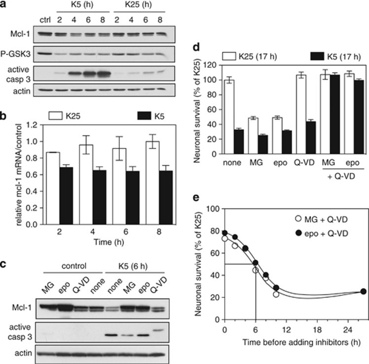
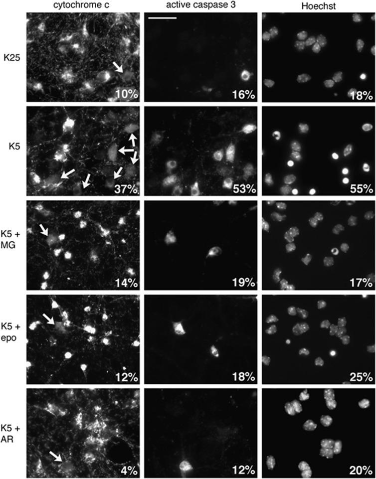
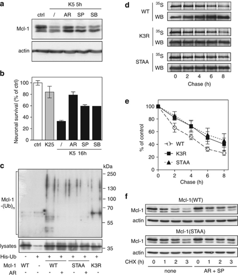
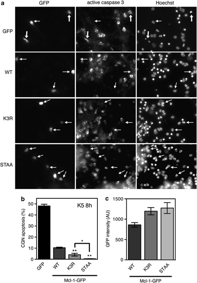
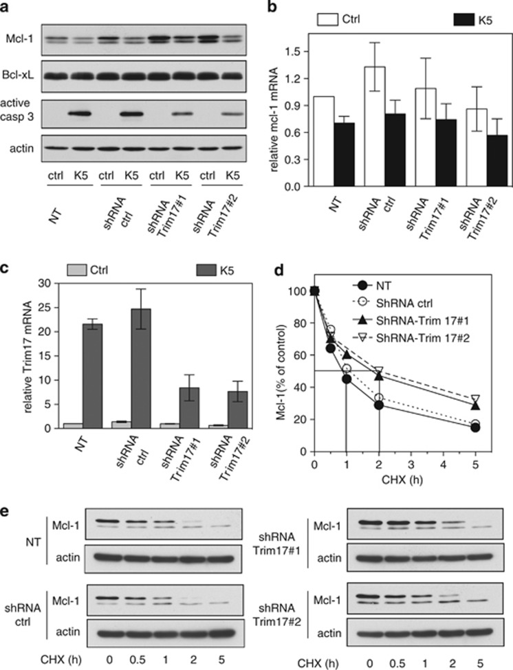
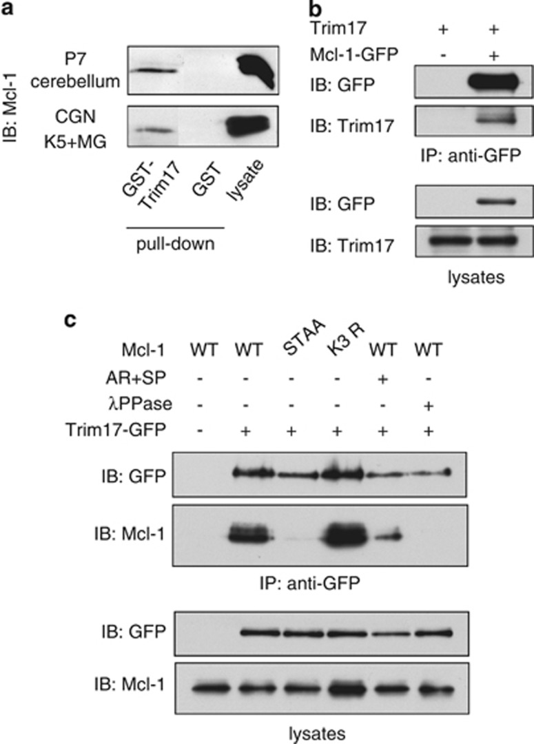
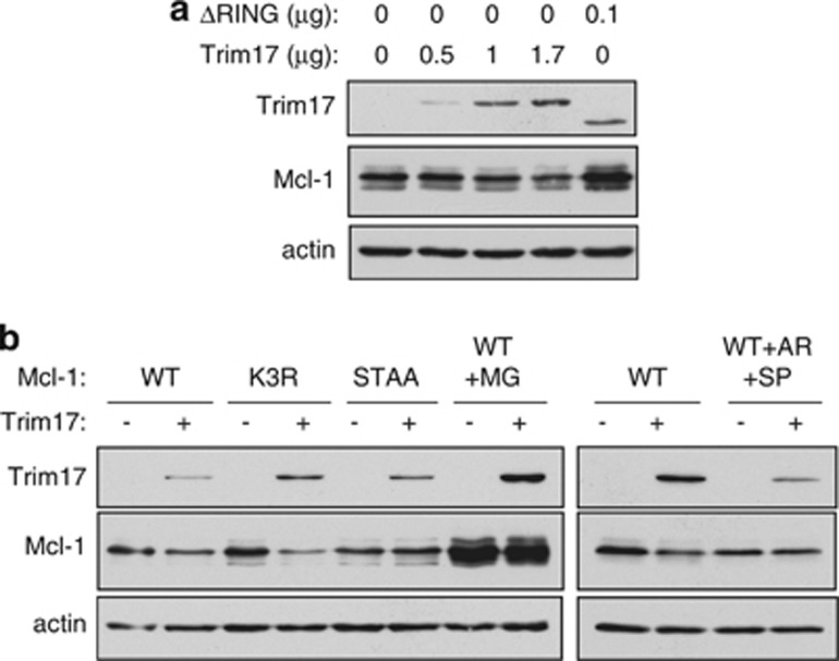
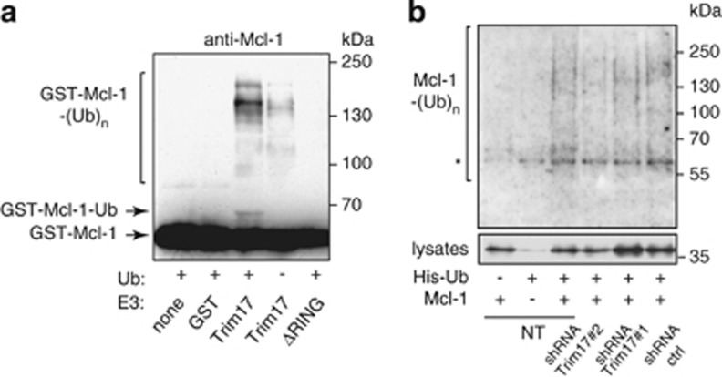
Short-term proteasome inhibition has been shown to prevent neuronal apoptosis. However, the key pro-survival proteins that must be degraded for triggering neuronal death are mostly unknown. Here, we show that Mcl-1, an anti-apoptotic Bcl-2 family member, is degraded by the proteasome during neuronal apoptosis. Using primary cultures of cerebellar granule neurons deprived of serum and KCl, we found that ubiquitination and proteasomal degradation of Mcl-1 depended on its prior phosphorylation by GSK3, providing the first insight into post-translational regulation of Mcl-1 in neurons. In a previous study, we have reported that the E3 ubiquitin-ligase Trim17 is both necessary and sufficient for neuronal apoptosis. Here, we identified Trim17 as a novel E3 ubiquitin-ligase for Mcl-1. Indeed, Trim17 co-immunoprecipitated with Mcl-1. Trim17 ubiquitinated Mcl-1 in vitro. Overexpression of Trim17 decreased the protein level of Mcl-1 in a phosphorylation- and proteasome-dependent manner. Finally, knock down of Trim17 expression reduced both ubiquitination and degradation of Mcl-1 in neurons. Moreover, impairment of Mcl-1 phosphorylation, by kinase inhibition or point mutations, not only decreased ubiquitination and degradation of Mcl-1, but also blocked the physical interaction between Trim17 and Mcl-1. As this stabilization of Mcl-1 increased its neuroprotective effect, our data strongly suggest that Trim17-mediated ubiquitination and degradation of Mcl-1 is necessary for initiating neuronal death.


Figures

Figure 1
Figure 1
Mcl-1 is degraded by the proteasome during KCl deprivation-induced apoptosis in CGNs. (a) CGN primary cultures were left untreated (ctrl) or washed and switched to serum free medium containing either 25 mM KCl (K25) or 5 mM KCl (K5) for increasing times. Total protein extracts were prepared and western blot analysis was performed with antibodies against Mcl-1, the phosphorylated form (Ser9) of GSK3, the active (cleaved) form of caspase 3 and actin (loading control). (b) CGNs were incubated in K25 or in K5 medium for the indicated times. Total RNA was extracted and mcl-1 mRNA levels were estimated by quantitative RT-PCR. Fold change was calculated by comparison with neurons maintained in the initial culture medium (ctrl). Data are means±S.D. of triplicates and are representative of five independent experiments. (c) CGNs were left untreated (control) or switched to K5 medium in the presence or absence of 20 μM MG-132 (MG), 10 μM epoxomicin (epo) or 100 μM Q-VD-OPh (Q-VD) for 6 h. Total protein extracts were analyzed by western blot using antibodies against Mcl-1, active caspase 3 and actin. (d) CGN primary cultures were switched to K25 or K5 medium in the presence or absence of 5 μM MG-132, 5 μM epoxomicin, 50 μM Q-VD-OPh or a combination of proteasome and caspase inhibitors for 17 h. Then, neuronal viability was determined by MTT assay. Data are the means±S.D. of triplicate determinations obtained in a typical experiment representative of five independent experiments. Results are expressed as the percentage of surviving neurons compared to neurons maintained in K25 medium. (e) To determine the proteasomal commitment point, CGNs were switched to K5 medium at time 0 to induce apoptosis. At increasing times after deprivation, proteasome inhibitors were added to the medium together with a caspase inhibitor: 5 μM MG-132+50 μM Q-VD-OPh (open circle) or 5 μM epoxomicin+50 μM Q-VD-OPh (closed circle), in order to rescue neurons. Neuronal viability for all conditions was estimated 27 h after KCl deprivation by MTT assay. Results are expressed as percentage of surviving neurons compared with cultures maintained in K25 throughout the experiment. Data are the means of two independent experiments performed with different neuronal cultures
Figure 2
Figure 2
Proteasome and GSK3 inhibitors protect neurons from apoptosis by preventing cytochrome c release and caspase activation. CGN primary cultures were washed and switched to serum-free medium containing either 25mM KCl (K25) or 5 mM KCl (K5) in the presence or absence of 20 μM MG-132 (MG), 10 μM epoxomicin (epo) or 10 μM AR-A014418 for 8 h. After fixation, nuclear condensation was visualized by Hoechst 33258 staining. Cytochrome c subcellular localization and caspase 3 activation were detected by immunofluorescence. In healthy neurons, cytochrome c immunostaining is intense and punctate both in cell bodies and in neurites (axons and dendrites), indicating mitochondrial localization. In apoptotic neurons, the staining is faint and diffuse, indicating that cytochrome c has been released from mitochondria. At late stages of apoptosis, the staining disappears because cytochrome c is rapidly degraded after release. The percentages of neurons with a condensed nucleus, showing a diffuse staining for cytochrome c or positive for active caspase 3 are indicated for each condition (500 neurons scored in a blinded manner for each condition). Arrows indicate cell bodies with a diffuse cytochrome c staining. Scale bar is 20 μm. Data are representative of three independent experiments
Figure 3
Figure 3
Prior phosphorylation of Mcl-1 is required for its ubiquitination and subsequent degradation. (a) CGNs were left untreated (ctrl) or washed and switched to K5 medium in the presence or absence of 10 μM AR-A014418, 10 μM SP600125 or 20 μM SB 203580 for 5 h. Total protein extracts were analyzed by immunoblotting using antibodies against Mcl-1 and actin. (b) CGN cultures were incubated in K25 or K5 medium in the presence or absence of 10 μM AR-A014418, 10 μM SP600125 or 20 μM SB 203580 for 16 h. Then, neuronal viability was determined by MTT assay. Data are the means±S.D. of triplicate determinations obtained in a typical experiment representative of three independent experiments. Results are expressed as the percentage of surviving neurons compared to neurons maintained in initial culture medium (ctrl). (c) CGNs were transfected with untagged Mcl-1(WT), Mcl-1(STAA) or Mcl-1(K3R), or with empty plasmid (−), together with His-tagged ubiquitin (+) or empty plasmid (−) for 18 h. Then, neurons were incubated for 6 h with 25 μM MG-132, in the presence or the absence of 10 μM AR-A011418 (AR). The ubiquitinated proteins were purified using nickel beads and analyzed by western blotting using anti-Mcl-1 antibody to detect ubiquitin-conjugated Mcl-1. In a separate SDS-PAGE, samples of the input lysates used for the purification were analyzed with anti-Mcl-1 antibody to estimate the expression level of the different forms of transfected Mcl-1. (d) CGNs were transfected with Mcl-1(WT)-GFP, Mcl-1(K3R)-GFP or Mcl-1(STAA)-GFP for 18 h. Then, neurons were metabolically labeled with [35S]-Met for 2 h (pulse) and harvested at different times after washing and incubation in K5 medium (chase). The different forms of Mcl-1-GFP were then immunoprecipitated using GFP-trap beads, separated by SDS-PAGE and visualized by both autoradiography (35S) and immunodetection of Mcl-1 (WB). Note that a higher amount of Mcl-1(WT) was immunoprecipitated compared to the mutants in the three last time points. This may give the feeling that it is more stable than it actually is. (e) The intensity of the bands on the autoradiograms and on the immunoblots of different experiments performed as in (d) was quantified. For each experiment, the radioactivity associated with the different forms of Mcl-1-GFP was normalized by the amount of protein immunoprecipitated in each condition and plotted against chase time. Data are the mean±S.E.M. of three independent experiments. (f) CGNs were transfected with untagged Mcl-1(WT) or Mcl-1(STAA). Eighteen hours after transfection, neurons were pre-incubated for 1 h in K5 medium in the presence or the absence of 10 μM AR-A011418 and 10 μM SP600125 (AR+SP). Then, 10 μg/ml cycloheximide (CHX) was added, and neurons were harvested after the indicated times after CHX addition. Proteins were analyzed by western blot using antibodies against Mcl-1 and actin
Figure 4
Figure 4
Neuroprotective effect of the different forms of Mcl-1. (a) CGN primary cultures were transfected with GFP (used as a negative control) or the different forms of Mcl-1 fused to GFP, for 16 h. Then, neurons were incubated in K5 medium for 8 h. Caspase 3 activation was determined by immunofluorescence and nuclear condensation was visualized by Hoechst staining. Thick arrows indicate GFP-expressing neurons that undergo apoptosis, whereas thin arrows indicate healthy neurons expressing GFP or the different forms of Mcl-1-GFP. (b) The percentage of apoptosis among transfected neurons was assessed by examining cell morphology and nuclear condensation of GFP-positive neurons. Data are the mean±S.D. of three independent experiments. **P<0.001 significantly different from apoptosis observed in neurons expressing Mcl-1(WT)-GFP (ANOVA followed by Student-Newman–Keuls multiple comparison test). The difference observed between neurons expressing Mcl-1(K3R)-GFP and Mcl-1(STAA)-GFP is also significant (*P<0.01 using the same statistical tests). (c) GFP intensity (AU: Arbitrary Unit) in the cell body of transfected neurons was measured using MetaMorph. Data are the mean±S.E.M. of about 100 neurons for each condition
Figure 5
Figure 5
Silencing of Trim17 favours Mcl-1 stabilization. (a) CGNs were left untreated (non-transduced: NT) or were transduced with lentiviral particles expressing shRNA sequences (one control and two against Trim17) one day after plating. At DIV 6, neurons were incubated for 6 h in K5 medium or maintained in the initial culture medium (ctrl). Then, proteins were analyzed by western blot using antibodies against Mcl-1, Bcl-x, active caspase 3 and actin. (b and c) CGNs were transduced or not as in (a). Then, neurons were incubated for 4 h in K5 medium or maintained in the initial culture medium (ctrl). Total RNA was extracted and the mRNA levels of mcl-1 (b) and Trim17 (c) were estimated under each condition by quantitative RT-PCR. Fold change was calculated by comparison with non-transduced neurons maintained in the initial culture medium. Data are means±s.d. of triplicate measurements of one experiment that is representative of three independent experiments. (d and e) CGNs were transduced or not as in (a) and incubated for increasing times with 10 μg/ml cycloheximide (CHX). Proteins were analyzed by western blot using antibodies against Mcl-1 and actin. (d) The intensity of the Mcl-1 bands presented in a representative experiment performed as in (e) was estimated and expressed as a percentage of the corresponding value for time zero
Figure 6
Figure 6
Trim17 interacts with phosphorylated Mcl-1. (a) GST pull-down assays were performed with lysates from P7 mice cerebella, or from CGNs incubated for 6 h in K5 medium with 20 μM MG-132, using recombinant GST-Trim17 or GST (as a negative control) purified on glutathione magnetic beads. Materials bound to recombinant proteins (pull-down) and input lysates were analyzed by western blot using anti-Mcl-1 antibody. (b) Neuro2A cells were transfected with Trim17 together with Mcl-1-GFP or GFP alone (as a negative control) for 24 h. Then, cells were treated for 8 h with 20 μM MG-132 before cell harvesting. Cell lysates were subjected to immunoprecipitation in the presence of phosphatase inhibitors using GFP-trap beads. Immunoprecipitates and lysates were analyzed by western blot using anti-GFP and anti-Trim17 antibodies. (c) Neuro2A cells were transfected with the different untagged variants of Mcl-1 together with Trim17-GFP or GFP alone for 24 h. Then, cells were treated for 8 h with 20 μM MG-132, in the absence or the presence of 10 μM AR-A014418 and 10 μM SP600125 (AR+SP) before cell harvesting. Cell lysates were subjected to immunoprecipitation as in (b). When indicated, beads were treated with λ-phosphatase (λPPase) after immunoprecipitation, and were further washed before elution. Immunoprecipitates and lysates were analyzed by western blot using anti-GFP and anti-Mcl-1 antibodies
Figure 7
Figure 7
Overexpressed Trim17 decreases the protein level of Mcl-1. (a) Neuro2A cells were transfected with 0.3 μg pCI-Mcl-1(WT) together with the indicated amounts of pCI-Trim17 or pCI-Trim17(ΔRING). Twenty four hours after transfection, cell lysates were analyzed by western blot using antibodies against Trim17, Mcl-1 and actin. (b) Neuro2A cells were transfected for 24 h with the different forms of Mcl-1 (0.5 μg of each plasmid), together with 1 μg pCI-Trim17 when indicated. Then, cells were treated for 5 h with 25 μM MG-132 (MG), or 10 μM AR-A014418 and 10 μM SP600125 (AR+SP), before cell harvesting, when indicated. Cell lysates were analyzed by western blot using antibodies against Trim17, Mcl-1 and actin
Figure 8
Figure 8
Trim17 promotes Mcl-1 ubiquitination both in vitro and in vivo. (a) Purified recombinant mouse Mcl-1 was first phosphorylated by recombinant JNK and GSK3 in vitro. Then, it was incubated for 2 h in the in vitro ubiquitination reaction mix with different recombinant proteins: GST, GST-Trim17(WT) or GST-Trim17(ΔRING), in the presence or the absence of ubiquitin, as indicated. Mcl-1 ubiquitination was examined by anti-Mcl-1 immunoblotting. The arrows indicate unmodified Mcl-1 (GST-Mcl-1) and mono- or di-ubiquitinated form of Mcl-1 (GST-Mcl-1-Ub). Higher bands are poly- or multi-ubiquitinated forms of Mcl-1 (GST-Mcl-1-(Ub)n). (b) CGNs were left untreated (non transduced: NT) or were transduced with lentiviral particles expressing shRNA sequences (one control and two against Trim17), one day after plating. At DIV 5, neurons were transfected with Mcl-1(WT), His-tagged ubiquitin or both for 18 h. The ubiquitinated proteins were purified using nickel beads, separated by SDS-PAGE and visualized by immunoblotting with anti-Mcl-1 antibody to detect ubiquitin-conjugated Mcl-1. In a separate SDS-PAGE, samples of the input lysates were analyzed with anti-Mcl-1 antibody to estimate the expression level of transfected Mcl-1 in the different conditions. Normalization of the ubiquitination signal by the expression level of Mcl-1 in lysates shows that ubiquitination is approximately reduced by half after transduction with both shRNA-Trim17#1 and #2 compared to shRNA ctrl. *Indicates a non-specific band

Similar articles

Cited by

References

    1. Yuan J, Yankner BA. Apoptosis in the nervous system. Nature. 2000;407:802–809. - PubMed
    1. Chipuk JE, Moldoveanu T, Llambi F, Parsons MJ, Green DR. The BCL-2 family reunion. Mol Cell. 2010;37:299–310. - PMC - PubMed
    1. Warr MR, Shore GC. Unique biology of Mcl-1: therapeutic opportunities in cancer. Curr Mol Med. 2008;8:138–147. - PubMed
    1. Nijhawan D, Fang M, Traer E, Zhong Q, Gao W, Du F, et al. Elimination of Mcl-1 is required for the initiation of apoptosis following ultraviolet irradiation. Genes Dev. 2003;17:1475–1486. - PMC - PubMed
    1. Ding Q, He X, Hsu JM, Xia W, Chen CT, Li LY, et al. Degradation of Mcl-1 by beta-TrCP mediates glycogen synthase kinase 3-induced tumor suppression and chemosensitization. Mol Cell Biol. 2007;27:4006–4017. - PMC - PubMed

Publication types

MeSH terms

Substances