. 2012 Nov;158(Pt 11):2753-2764.
doi: 10.1099/mic.0.062547-0. Epub 2012 Sep 13.

Substitutions in the Escherichia coli RNA polymerase inhibitor T7 Gp2 that allow inhibition of transcription when the primary interaction interface between Gp2 and RNA polymerase becomes compromised

Affiliations

Substitutions in the Escherichia coli RNA polymerase inhibitor T7 Gp2 that allow inhibition of transcription when the primary interaction interface between Gp2 and RNA polymerase becomes compromised

Andrey Shadrin et al. Microbiology (Reading). 2012 Nov.

Abstract

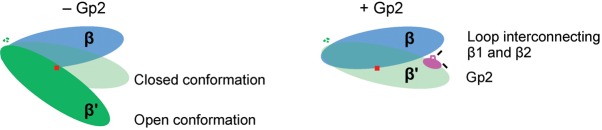
The Escherichia coli-infecting bacteriophage T7 encodes a 7 kDa protein, called Gp2, which is a potent inhibitor of the host RNA polymerase (RNAp). Gp2 is essential for T7 phage development. The interaction site for Gp2 on the E. coli RNAp is the β' jaw domain, which is part of the DNA binding channel. The binding of Gp2 to the β' jaw antagonizes several steps associated with interactions between the RNAp and promoter DNA, leading to inhibition of transcription at the open promoter complex formation step. In the structure of the complex formed between Gp2 and a fragment of the β' jaw, amino acid residues in the β3 strand of Gp2 contribute to the primary interaction interface with the β' jaw. The 7009 E. coli strain is resistant to T7 because it carries a charge reversal point mutation in the β' jaw that prevents Gp2 binding. However, a T7 phage encoding a mutant form of Gp2, called Gp2(β), which carries triple amino acid substitutions E24K, F27Y and R56C, can productively infect this strain. By studying the molecular basis of inhibition of RNAp from the 7009 strain by Gp2(β), we provide several lines of evidence that the E24K and F27Y substitutions facilitate an interaction with RNAp when the primary interaction interface with the β' jaw is compromised. The proposed additional interaction interface between RNAp and Gp2 may contribute to the multipronged mechanism of transcription inhibition by Gp2.


Figures

Fig. 1.
Fig. 1.
Sequence alignment analysis of T7 Gp2-like proteins. (a) Ribbon representation of the T7 Gp2–E. coli RNAp β′ jaw domain fragment (residues 1153–1213) complex. Gp2 is shown in cyan and the β′ jaw domain fragment is shown in green. The positions of the amino acids E24, F27 and R56 in T7 Gp2 are indicated along with the E1158 and E1188 residues in the β′ jaw domain. (b) Alignment of amino acid sequences from known Gp2-like proteins. The sequences are displayed in single-letter amino acid code, with the length of the sequence indicated on the far right. The localization of the β-strands and α-helix based on the structure of T7 Gp2 is indicated. The highly conserved R56 and R58 residues are highlighted in blue. The positions of amino acids residues E24 and F27 in T7 Gp2 are underlined and in bold type. The arginine residue, corresponding to T7 Gp2 amino acid residue S23 in Gp2-like proteins in phages CR8, CR44b, K1F and EcoDS1, is also underlined in bold type. (c) Alignment of the β′ jaw domain amino acid residues 1153–1213 from a representative set of host RNAps, with those infected by T7-like phages encoding Gp2-like proteins indicated by asterisks. Amino acid residues corresponding to E1158 and E1188 of the E. coli RNAp are shown in bold type. (d) As in (a), except that the structural models of Gp2-like proteins CR44b_13 (orange) and CR8_16 (pink) are superimposed and amino acid residue R15 is indicated. The structural models of the Gp2 homologues were calculated with swiss-model using NMR structures of T7 Gp2 as a template (Arnold et al., 2006; Cámara et al., 2010).
Fig. 2.
Fig. 2.
Establishing a simple in vivo assay to measure the activity of Gp2 in the absence of T7 phage infection. Growth curves of (a) E. coli MG1655 and (b) 7009 cells expressing Gp2 upon induction with arabinose at t = 0. The solid red, dotted and solid black lines represent cells harbouring pCS1 : Gp2WT, pBAD18 (empty vector) and pCS1 : Gp2R56E, respectively; the dashed line represents cells harbouring pCS1 : Gp2WT to which ampicillin was not added to the growth medium. (c) As in (a), except that at t = 2 h, the cells were pelleted and resuspended in fresh LB to remove inducer (arabinose) prior to continuation of growth. The dashed red line represents cells that were induced for the second time (see text for details) at t = 6 h. (d) Quantification of Gp2 molecules per E. coli MG1655 cell at t = 0.5, 1 and 2 h post induction of Gp2. Top panel: digitalized image of the Western blot probed with HRP-conjugated anti-His monoclonal antibodies containing purified Gp2 (as a reference standard) and whole-cell protein extracts corresponding to 2.5×10⁷ cells. Bottom-left panel: calibration curve relating signal intensity to the amount of purified Gp2 (in ng). Bottom-right panel: table showing the calculated number of molecules of Gp2 per cell for each time point sampled post induction.
Fig. 3.
Fig. 3.
Ability of single, double and triple mutant versions of Gp2 based on Gp2β to attenuate bacterial growth upon induction. Growth curves of (a) E. coli MG1655, (b) 7009 and (c) BR3 expressing wild-type Gp2 and Gp2 mutants containing various combinations of mutations based on Gp2β. Gp2 expression was induced with arabinose at t = 0. In each graph, the solid red, dotted and dashed black lines represent cells harbouring pCS1 : Gp2WT, pBAD18 (empty vector) and pCS1 : Gp2 encoding the indicated mutant form of Gp2, respectively. In panels (a–c), the same relevant traces for cells harbouring pCS1 : Gp2WT and pBAD18 (empty vector) were used.
Fig. 4.
Fig. 4.
Amino acids 14–59 of Gp2 are sufficient to inhibit the E. coli RNAp. Growth curves of E. coli MG1655 cells in which the expression of (a) Gp214–59, (b) Gp214–59/R56C and (c) Gp214–59/E24K/F27Y was induced at t = 0 (dashed lines). In each graph, the solid red and dotted lines represent cells harbouring pCS1 : Gp2WT and pBAD18 (empty vector), respectively.
Fig. 5.
Fig. 5.
Experiments with Gp2-like proteins from C. rodentium phages CR44b and CR8. Growth curves of (a) E. coli MG1655, (b) JE1134, (c) 7009 and (d) BR3 cells in which the expression of T7 Gp2 and Gp2-like proteins CR44b_13 and CR8_16 was induced at t = 0 (solid red lines). In each graph, the dotted line represents cells harbouring pBAD18 (empty vector). (e) Autoradiograph of a 20 % (w/v) denaturing gel showing synthesis of the transcript ApApUpU (the 32P-labelled nucleotides underlined) from the lacUV5 promoter by the σ70-containing RNAp in the presence of Gp2, CR44b_13 and CR8_16. The molar ratio of RNAP to Gp2/Gp2-like proteins is indicated above each lane. The percentage of ApApUpU transcript synthesized (%A) by the RNAp is calculated relative to the reaction without Gp2 and is indicated at the bottom of each lane. (f) Growth curves of E. coli MG1655 (left) and BR3 (right) cells expressing CR44b_13 and harbouring the R15E amino acid substitution. Induction with arabinose was performed at t = 0.
Fig. 6.
Fig. 6.
Gp2 induces RNAp to adopt the ‘closed state’ conformation. Cartoon showing how the region including and surrounding the β1 and β2 strands in Gp2 could contribute to inducing the closed state conformation in the RNAp.

Similar articles

Cited by

References

    1. Arnold K., Bordoli L., Kopp J., Schwede T. (2006). The swiss-model workspace: a web-based environment for protein structure homology modelling. Bioinformatics 22, 195–201. 10.1093/bioinformatics/bti770 - DOI - PubMed
    1. Belogurov G. A., Vassylyeva M. N., Svetlov V., Klyuyev S., Grishin N. V., Vassylyev D. G., Artsimovitch I. (2007). Structural basis for converting a general transcription factor into an operon-specific virulence regulator. Mol Cell 26, 117–129. 10.1016/j.molcel.2007.02.021 - DOI - PMC - PubMed
    1. Browning D. F., Busby S. J. (2004). The regulation of bacterial transcription initiation. Nat Rev Microbiol 2, 57–65. 10.1038/nrmicro787 - DOI - PubMed
    1. Burck K. B., Miller R. C., Jr (1978). Marker rescue and partial replication of bacteriophage T7 DNA. Proc Natl Acad Sci U S A 75, 6144–6148. 10.1073/pnas.75.12.6144 - DOI - PMC - PubMed
    1. Cámara B., Liu M., Reynolds J., Shadrin A., Liu B., Kwok K., Simpson P., Weinzierl R., Severinov K. & other authors (2010). T7 phage protein Gp2 inhibits the Escherichia coli RNA polymerase by antagonizing stable DNA strand separation near the transcription start site. Proc Natl Acad Sci U S A 107, 2247–2252. 10.1073/pnas.0907908107 - DOI - PMC - PubMed

Publication types

MeSH terms