

Comparison of velocity vector imaging echocardiography with magnetic resonance imaging in mouse models of cardiomyopathy
- PMID: 22977126
- PMCID: PMC3504170
- DOI: 10.1161/CIRCIMAGING.111.972406
Comparison of velocity vector imaging echocardiography with magnetic resonance imaging in mouse models of cardiomyopathy
Abstract
Background: Myocardial strain imaging using echocardiography can be a cost-effective method to quantify ventricular wall motion objectively, but few studies have compared strain measured with echocardiography against magnetic resonance imaging (MRI) in small animals.
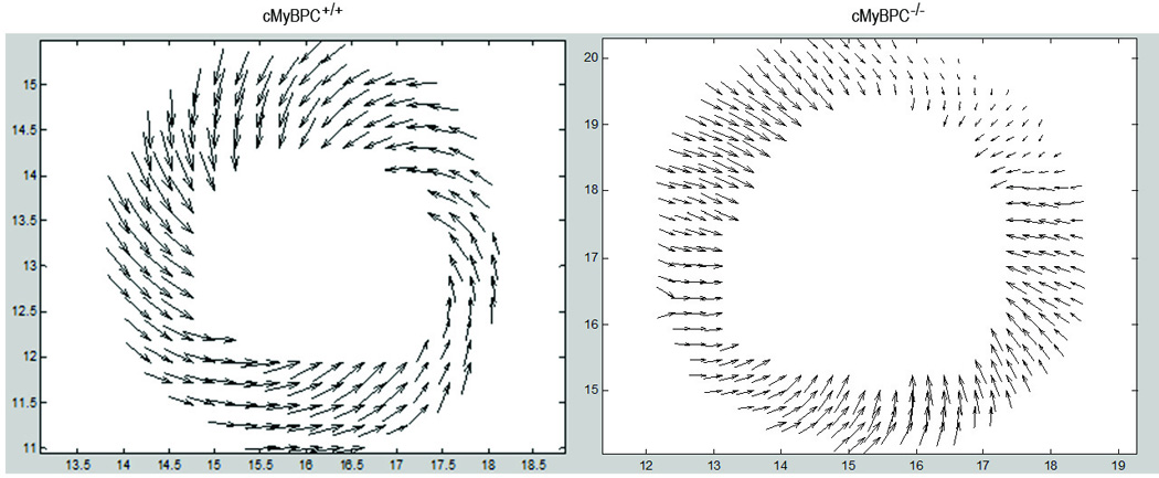
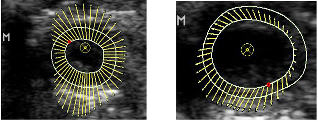
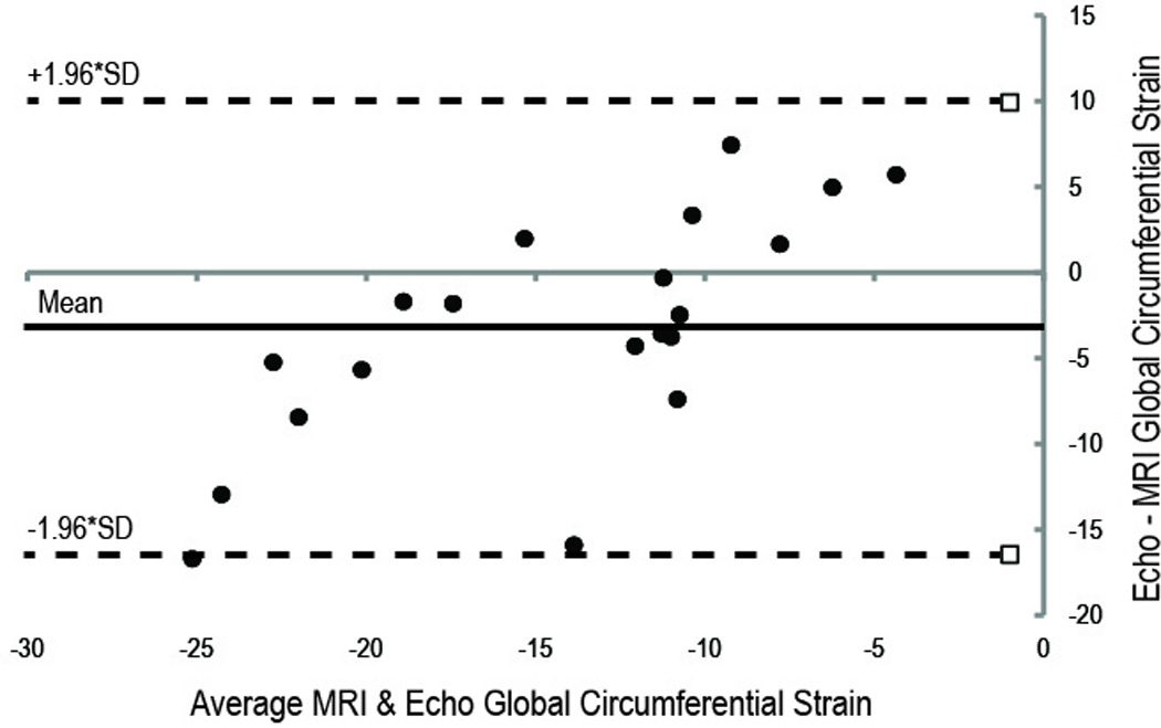
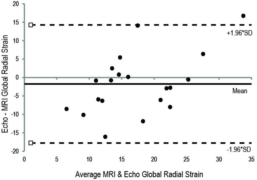
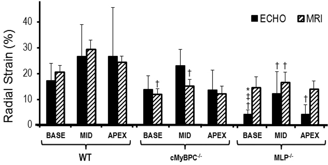
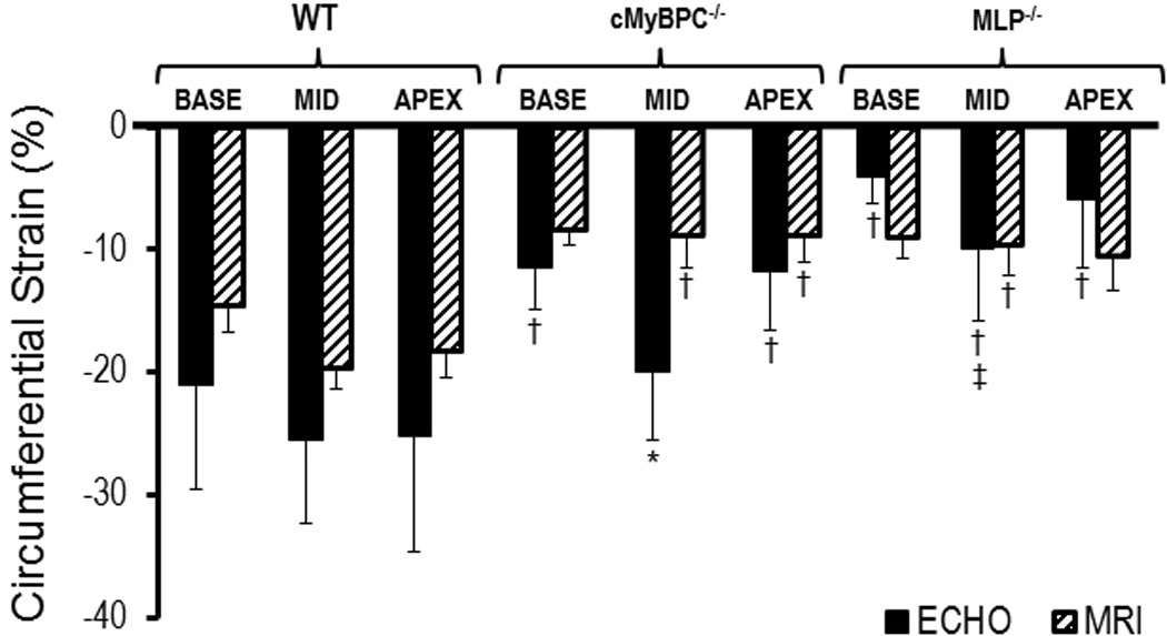
Methods and results: We compared circumferential strain (CS) and radial strain (RS) measured with echocardiography (velocity vector imaging [VVI]) to displacement encoding with stimulated-echo MRI in 2 mouse models of cardiomyopathy. In 3-month-old mice with gene targeted deficiency of cardiac myosin-binding protein-C (cMyBP-C(-/-), n=6) or muscle LIM protein (MLP(-/-), n=6), and wild-type mice (n=8), myocardial strains were measured at 3 cross-sectional levels and averaged to obtain global strains. There was modest correlation between VVI and MRI measured strains, with global CS yielding stronger correlation compared with global RS (CS R(2)=0.4452 versus RS R(2)=0.2794, both P<0.05). Overall, strain measured by VVI was more variable than MRI (P<0.05) and the limits of agreement were slightly, but not significantly (P=0.14), closer for global CS than RS. Both VVI and MRI strain measurements showed significantly lower global CS strain in the knockout groups compared with the wild type. The VVI (but not MRI) CS strain measurements were different between the 2 knockout groups (-14.5±3.8% versus -6.6±4.0%, cMyBP-C(-/-) versus MLP(-/-) respectively, P<0.05).
Conclusions: Measurements of left ventricular CS and RS are feasible in small animals using 2-dimensional echocardiography. VVI and MRI strain measurements correlated modestly and the agreement between the modalities tended to be greater for CS than RS. Although VVI and MRI strains were able to differentiate between wild-type and knockout mice, only global CS VVI differentiated between the 2 models of cardiomyopathy.
Figures
References
-
- Popovic ZB, Benejam C, Bian J, Mal N, Drinko J, Lee K, Forudi F, Reeg R, Greenberg NL, Thomas JD, Penn MS. Speckle-tracking echocardiography correctly identifies segmental left ventricular dysfunction induced by scarring in a rat model of myocardial infarction. Am J Physiol Heart Circ Physiol. 2007;292:H2809–H2816. - PubMed
-
- Bachner-Hinenzon N, Ertracht O, Leitman M, Vered Z, Shimoni S, Beeri R, Binah O, Adam D. Layer-specific strain analysis by speckle tracking echocardiography reveals differences in left ventricular function between rats and humans. AJP: Heart and Circulatory Physiology. 2010;299:H664–H672. - PubMed
-
- Hoit BD. Echocardiographic characterization of the cardiovascular phenotype in rodent models. Toxicol Pathol. 2006;34:105–110. - PubMed
-
- Marwick TH, Raman SV, Carrio I, Bax JJ. Recent developments in heart failure imaging. JACC Cardiovasc Imaging. 2010;3:429–439. - PubMed
-
- Borg AN, Ray SG. A unifying framework for understanding heart failure? Response to"left ventricular torsion by two-dimensional speckle tracking echocardiography in patients with diastolic dysfunction and normal ejection fraction" by park sj et al. J Am Soc Echocardiogr. 2009;22:318–320. author reply 321-312. - PubMed
Publication types
MeSH terms
Grants and funding
LinkOut - more resources
Full Text Sources
Medical
