. 2012 Oct 26;287(44):36634-8.
doi: 10.1074/jbc.C112.406769. Epub 2012 Sep 12.

Membrane tethering and nucleotide-dependent conformational changes drive mitochondrial genome maintenance (Mgm1) protein-mediated membrane fusion

Affiliations

Membrane tethering and nucleotide-dependent conformational changes drive mitochondrial genome maintenance (Mgm1) protein-mediated membrane fusion

Inbal Abutbul-Ionita et al. J Biol Chem. .

Abstract

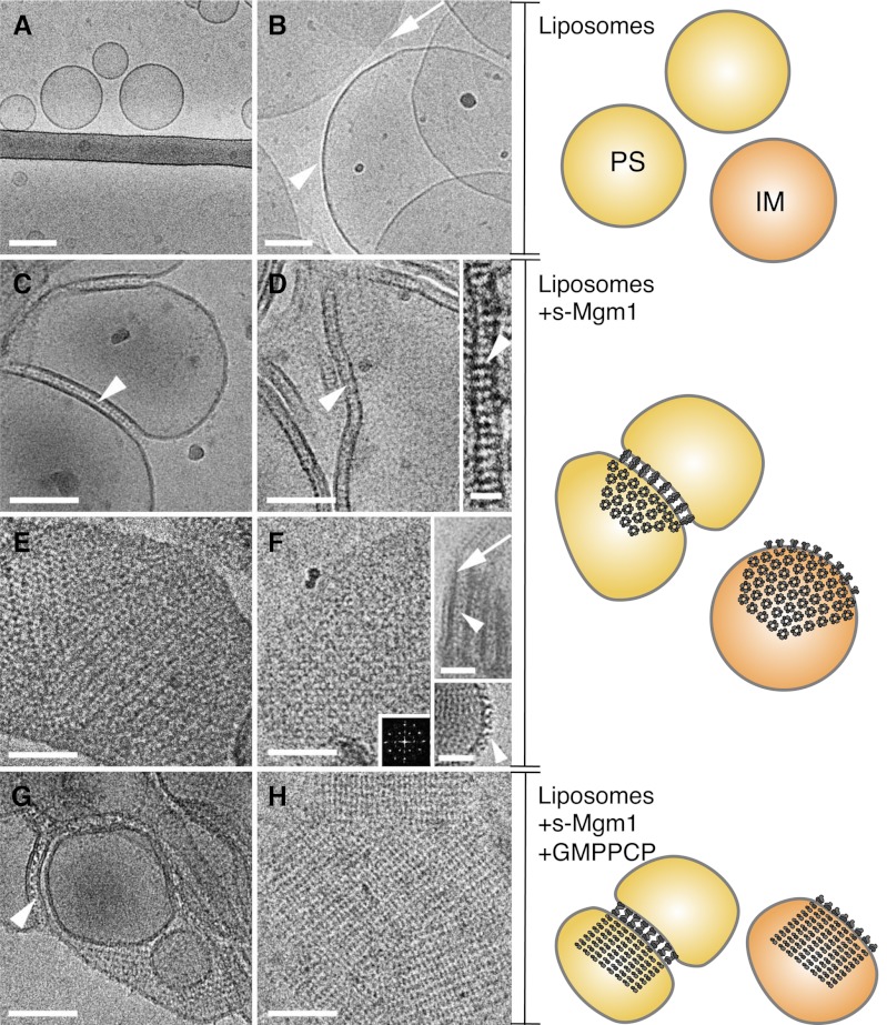
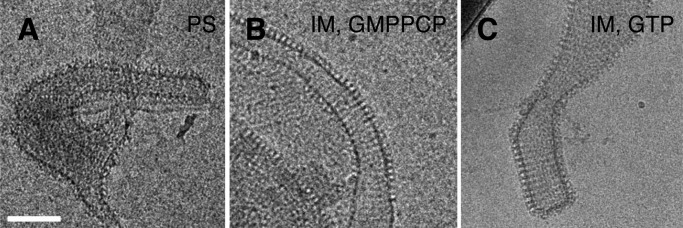
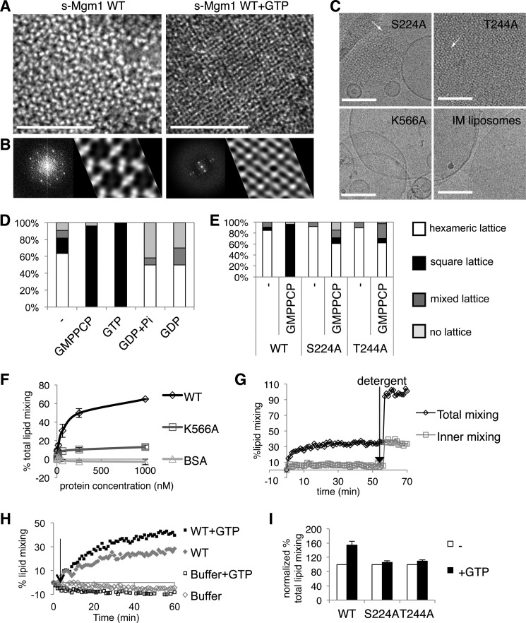
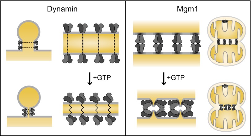
Cellular membrane remodeling events such as mitochondrial dynamics, vesicle budding, and cell division rely on the large GTPases of the dynamin superfamily. Dynamins have long been characterized as fission molecules; however, how they mediate membrane fusion is largely unknown. Here we have characterized by cryo-electron microscopy and in vitro liposome fusion assays how the mitochondrial dynamin Mgm1 may mediate membrane fusion. Using cryo-EM, we first demonstrate that the Mgm1 complex is able to tether opposing membranes to a gap of ∼15 nm, the size of mitochondrial cristae folds. We further show that the Mgm1 oligomer undergoes a dramatic GTP-dependent conformational change suggesting that s-Mgm1 interactions could overcome repelling forces at fusion sites and that ultrastructural changes could promote the fusion of opposing membranes. Together our findings provide mechanistic details of the two known in vivo functions of Mgm1, membrane fusion and cristae maintenance, and more generally shed light onto how dynamins may function as fusion proteins.


Figures

FIGURE 1.
FIGURE 1.
s-Mgm1 assembles onto and tethers liposomes. A and B, cryo-EM images of PS and IM liposomes, respectively. Arrowhead in B points to round liposome, and arrow points to the border of flat IM membrane. C and D, cryo-EM images show tethered PS liposomes and protein bridges (arrowheads). E and F, cryo-EM images show a crystalline protein array on PS (E) and IM (F) liposomes. The diffraction spots in F show the three-fold lattice symmetry. Arrowhead and arrows (E, upper inset) mark protein bridges and lipid bilayer. Lower inset shows T-shaped s-Mgm1 structures (arrowhead) on the outer liposome surface. G and H, cryo-EM images of crystalline arrays of s-Mgm1 in the presence of GMPPCP on PS (G) and IM (H) liposomes. Scale bar is 100 nm (A–H), 25 nm (inset in D), and 50 nm (insets in F). Schematics on the right summarize the observations from the cryo-EM images on the left. Yellow and orange circles represent PS and IM liposomes, respectively. At the periphery of the liposomes, a pair of light and dark gray structures represents the side view of an s-Mgm1 dimer. The ring of light and dark gray structures represents the top view of s-Mgm1 oligomers. Upon the addition of GMPPCP (from middle to bottom panels), the hexameric array of s-Mgm1 reorganizes into a square array.
FIGURE 2.
FIGURE 2.
s-Mgm1 can transform liposomes into protein-decorated tubes. Tubes are created with both PS (A) and IM (B and C) liposomes, without and with nucleotides as indicated. s-Mgm1 and lipid concentrations are 0.7 and 0.45 mg/ml respectively. Scale bar is 100 nm.
FIGURE 3.
FIGURE 3.
s-Mgm1 oligomeric arrays undergo a nucleotide-dependent structural transition that enhances s-Mgm1 membrane fusion activity. A, EM images of s-Mgm1 arrays on IM liposomes in the presence (right) and absence (left) of GTP. B, corresponding two-dimensional crystal analysis on protein crystalline arrays. Scale bar is 100 nm. Two-dimensional Fourier transform analysis and projection maps show the average patterns of s-Mgm1 arrays. C, cryo-EM images of GTPase mutants, S224A and T244A, and a lipid-binding mutant K566A. Upper panels: cryo-EM images of ordered protein arrays on IM liposome formed by S224A (left) and T244A (right). Arrows mark hexameric arrays. Lower panels: K566A does not assemble onto liposomes nor alter their morphology (compare left and right images). Scale bar is 100 nm. D, quantification of the nucleotide-dependent structural transition as categorized into four lattices, hexameric, square, mixed, or none. s-Mgm1-bound IM liposomes were incubated with buffer containing no nucleotide or 2 mm nucleotide as indicated. E, quantification of wild-type, S224A, and T244A s-Mgm1 assembly onto IM liposomes, categorized into four lattices. Analysis shows a nucleotide-induced transition from hexameric to square lattice. F, NBD-rhodamine assay showing the lipid mixing activity of Mgm1. Wild-type s-Mgm1 causes lipid mixing in a concentration-dependent manner, whereas the lipid mixing activity of the lipid-binding mutant K566A is significantly impaired. No activity is found for the control (bovine serum albumin, BSA). G, the lipid mixing of the inner leaflet only by s-Mgm1 was monitored by dithionite treatment. s-Mgm1 lipid mixing activity was monitored by the fusion of dithionite-treated and untreated labeled liposomes indicative of the mixing of the inner leaflet phospholipids only and total phospholipids, respectively. Detergent was added to determine the maximal NBD signals. H, GTP enhances Mgm1 lipid mixing activity. Arrow points to the 3-min point after the fusion has been initiated. GTP (1 mm) increases the total lipid mixing by wild-type s-Mgm1 (0.125 μm). I, the bar graph shows that GTP addition increased total lipid mixing induced by wild-type s-Mgm1 but not by the GTPase mutants S224A and T244A. Three separate experiments were performed. Basal levels of lipid mixing were normalized to 100%. The error bars represent standard deviations.
FIGURE 4.
FIGURE 4.
Models of Mgm1-mediated membrane fusion. A model depicting the function of dynamin in fission (left), and s-Mgm1 in fusion (right). Both dynamin and Mgm1 shape membranes in vivo and in vitro. Dynamin dimers assemble into a helical collar, which upon GTPase activity constrict the underlying membrane to mediate fission. We propose that s-Mgm1 forms a homo-oligomeric complex in trans to create protein bridges and ordered lattices that tether opposing membranes to support mitochondrial inner membrane cristae structures, and to also undergo a GTP-induced transition to promote fusion.

References

    1. Danino D., Hinshaw J. E. (2001) Dynamin family of mechanoenzymes. Curr. Opin. Cell Biol. 13, 454–460 - PubMed
    1. Praefcke G. J., McMahon H. T. (2004) The dynamin superfamily: universal membrane tubulation and fission molecules? Nat. Rev. Mol. Cell Biol. 5, 133–147 - PubMed
    1. Takei K., Haucke V., Slepnev V., Farsad K., Salazar M., Chen H., De Camilli P. (1998) Generation of coated intermediates of clathrin-mediated endocytosis on protein-free liposomes. Cell 94, 131–141 - PubMed
    1. Sweitzer S. M., Hinshaw J. E. (1998) Dynamin undergoes a GTP-dependent conformational change causing vesiculation. Cell 93, 1021–1029 - PubMed
    1. Stowell M. H., Marks B., Wigge P., McMahon H. T. (1999) Nucleotide-dependent conformational changes in dynamin: evidence for a mechanochemical molecular spring. Nat. Cell Biol. 1, 27–32 - PubMed

Publication types

MeSH terms