. 2012 Oct 26;287(44):37395-405.
doi: 10.1074/jbc.M112.406439. Epub 2012 Sep 12.

Functional and structural characterization of PaeM, a colicin M-like bacteriocin produced by Pseudomonas aeruginosa

Affiliations

Functional and structural characterization of PaeM, a colicin M-like bacteriocin produced by Pseudomonas aeruginosa

Hélène Barreteau et al. J Biol Chem. .

Abstract

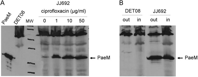
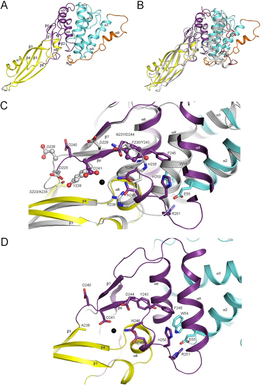
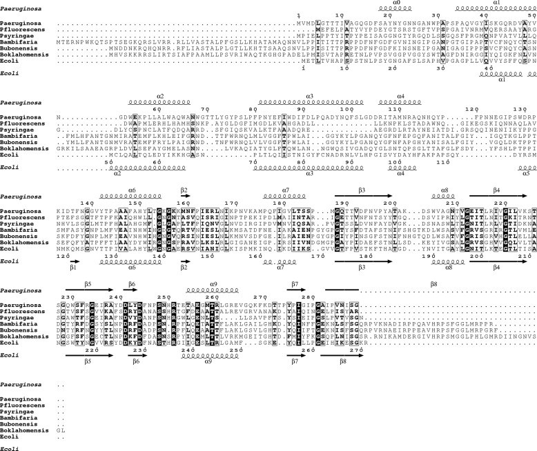
Colicin M (ColM) is the only enzymatic colicin reported to date that inhibits cell wall peptidoglycan biosynthesis. It catalyzes the specific degradation of the lipid intermediates involved in this pathway, thereby provoking lysis of susceptible Escherichia coli cells. A gene encoding a homologue of ColM was detected within the exoU-containing genomic island A carried by certain pathogenic Pseudomonas aeruginosa strains. This bacteriocin (pyocin) that we have named PaeM was crystallized, and its structure with and without an Mg(2+) ion bound was solved. In parallel, site-directed mutagenesis of conserved PaeM residues from the C-terminal domain was performed, confirming their essentiality for the protein activity both in vitro (lipid II-degrading activity) and in vivo (cytotoxicity against a susceptible P. aeruginosa strain). Although PaeM is structurally similar to ColM, the conformation of their active sites differs radically; in PaeM, residues essential for enzymatic activity and cytotoxicity converge toward a same pocket, whereas in ColM they are spread along a particularly elongated active site. We have also isolated a minimal domain corresponding to the C-terminal half of the PaeM protein and exhibiting a 70-fold higher enzymatic activity as compared with the full-length protein. This isolated domain of the PaeM bacteriocin was further shown to kill E. coli cells when addressed to the periplasm of these bacteria.


Figures

FIGURE 1.
FIGURE 1.
Production and excretion of PaeM. A, the P. aeruginosa JJ692 strain was grown and treated for 2 h by ciprofloxacin at the indicated concentrations. Crude cell protein extracts were prepared and analyzed by SDS-PAGE, and the PaeM protein was detected by Western-blot, as detailed in “Experimental Procedures.” Purified PaeM protein and a crude extract from strain DET08 were also analyzed as controls (two lanes on the left). B, the P. aeruginosa DET08 (PaeM-susceptible) and JJ692 (PaeM-producing) strains were grown and treated for 2 h by ciprofloxacin at 10 μg/ml. Cells were harvested, and the presence of the PaeM protein was searched for both in the external growth medium (out) and crude cell extracts (in) fractions.
FIGURE 2.
FIGURE 2.
Structure of PaeM. A, shown is a ribbon representation of the PaeM structure. The translocation and receptor binding domains are colored in orange and cyan, respectively. The catalytic domain is colored in yellow with the small subdomain in purple. The part of the His tag present at the C-terminal end of PaeM and defined in the 2 FoFc electron density map is shown in gray. B, superimposition of ColM onto PaeM is shown. PaeM is colored according to the same color code as panel A. The ColM translocation domain is shown in red, and the remaining domains are in gray. C, shown is a comparison of PaeM (same color code as panel A) and ColM (gray) active sites. Residues discussed in the manuscript are shown as sticks and/or ball and sticks. Residues and secondary structure elements from ColM are labeled in italics. The Mg2+ ion is depicted as a black sphere. D, shown is a detailed representation of the PaeM active site (same color code as panel C).
FIGURE 3.
FIGURE 3.
Sequence alignment of PaeM orthologues. Strictly conserved residues are in white on a black background. Partially conserved amino acids are boxed. Secondary structure elements present in the crystal structures of PaeM and ColM are shown above and below the alignment, respectively. The aligned PaeM orthologues sequences are from Pseudomonas aeruginosa (Paeruginosa), Pseudomonas fluorescens (Pfluorescens), Pseudomonas syringae (Psyringae), Burkholderia ambifaria (Bambifaria), Burkholderia ubonensis (Bubonensis), Burkholderia oklahomensis (Boklahomensis), and Escherichia coli (Ecoli).

References

    1. Cascales E., Buchanan S. K., Duché D., Kleanthous C., Lloubès R., Postle K., Riley M., Slatin S., Cavard D. (2007) Colicin biology. Microbiol. Mol. Biol. Rev. 71, 158–229 - PMC - PubMed
    1. Pugsley A. P. (1984) The ins and outs of colicins. Part II. Lethal action, immunity, and ecological implications. Microbiol. Sci. 1, 203–205 - PubMed
    1. Pugsley A. P. (1984) The ins and outs of colicins. Part I. Production and translocation across membranes. Microbiol. Sci. 1, 168–175 - PubMed
    1. Schaller K., Höltje J. V., Braun V. (1982) Colicin M is an inhibitor of murein biosynthesis. J. Bacteriol. 152, 994–1000 - PMC - PubMed
    1. Barreteau H., El Ghachi M., Barnéoud-Arnoulet A., Sacco E., Touzé T., Duché D., Gérard F., Brooks M., Patin D., Bouhss A., Blanot D., van Tilbeurgh H., Arthur M., Lloubès R., Mengin-Lecreulx D. (2012) Characterization of colicin M and its orthologs targeting bacterial cell wall peptidoglycan biosynthesis. Microb. Drug Resist. 18, 222–229 - PubMed

Publication types

MeSH terms

LinkOut - more resources