

Determination of optimal imaging mode for ultrasonographic detection of subdermal contraceptive rods: comparison of spatial compound, conventional, and tissue harmonic imaging methods
- PMID: 22977328
- PMCID: PMC3435858
- DOI: 10.3348/kjr.2012.13.5.602
Determination of optimal imaging mode for ultrasonographic detection of subdermal contraceptive rods: comparison of spatial compound, conventional, and tissue harmonic imaging methods
Abstract
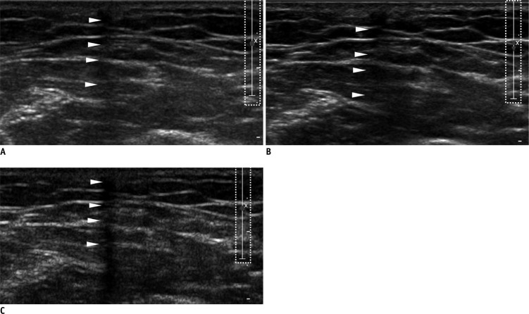
Objective: To determine which mode of ultrasonography (US), among the conventional, spatial compound, and tissue-harmonic methods, exhibits the best performance for the detection of Implanon® with respect to generation of posterior acoustic shadowing (PAS).
Materials and methods: A total of 21 patients, referred for localization of impalpable Implanon®, underwent US, using the three modes with default settings (i.e., wide focal zone). Representative transverse images of the rods, according to each mode for all patients, were obtained. The resulting 63 images were reviewed by four observers. The observers provided a confidence score for the presence of PAS, using a five-point scale ranging from 1 (definitely absent) to 5 (definitely present), with scores of 4 or 5 for PAS being considered as detection. The average scores of PAS, obtained from the three different modes for each observer, were compared using one-way repeated measure ANOVA. The detection rates were compared using a weighted least square method.
Results: Statistically, the tissue harmonic mode was significantly superior to the other two modes, when comparing the average scores of PAS for all observers (p < 0.00-1). The detection rate was also highest for the tissue harmonic mode (p < 0.001).
Conclusion: Tissue harmonic mode in uS appears to be the most suitable in detecting subdermal contraceptive implant rods.
Keywords: Implanon; Spatial compound; Subdermal contraceptive; Tissue harmonic; Ultrasonography; Wide focal zone.
Figures


References
-
- Shulman LP, Gabriel H. Management and localization strategies for the nonpalpable Implanon rod. Contraception. 2006;73:325–330. - PubMed
-
- Mascarenhas L. Insertion and removal of Implanon. Contraception. 1998;58(6 Suppl):79S–83S. - PubMed
-
- James P, Trenery J. Ultrasound localisation and removal of non-palpable Implanon implants. Aust N Z J Obstet Gynaecol. 2006;46:225–228. - PubMed
-
- Persaud T, Walling M, Geoghegan T, Buckley O, Stunell H, Torreggiani WC. Ultrasound-guided removal of Implanon devices. Eur Radiol. 2008;18:2582–2585. - PubMed
-
- McNeill G, Ward E, Halpenny D, Snow A, Torreggiani W. Ultrasound appearances of Implanon implanted contraceptive devices. JBR-BTR. 2009;92:259–260. - PubMed
Publication types
MeSH terms
Substances
LinkOut - more resources
Full Text Sources
Medical
