. 2011 May;2(3):491-497.
doi: 10.3892/etm.2011.242. Epub 2011 Mar 21.

Retinoid acid receptors in human colorectal cancer: An unexpected link with patient outcome

Affiliations

Retinoid acid receptors in human colorectal cancer: An unexpected link with patient outcome

Aurélie Perraud et al. Exp Ther Med. 2011 May.

Abstract

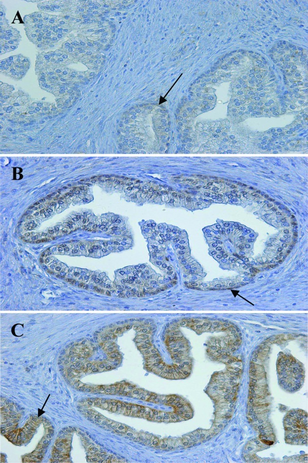
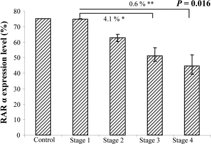
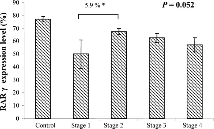
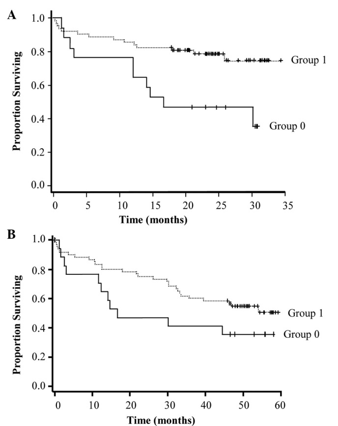
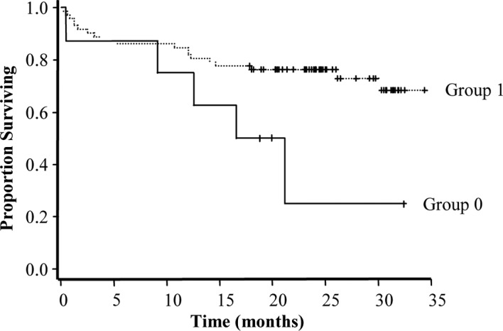
The status of the three retinoic acid receptors (RARs) α, β and γ in human colorectal cancer (CRC) has not as yet been examined. RARs are in part responsible for the actions of the retinoids (vitamin A and its derivatives), which are essential for human health and survival due to their extensive involvement in numerous cellular processes, in particular in epithelial morphology. The present study examined the expression of the three RARs in CRC using immunohistochemical analysis of paraffin-embedded tissue sections. RAR expression in tumor (T) and adjacent non-tumor (NT) specimens from stage I (n=6), stage II (n=34), stage III (n=26) and stage IV (n=14) CRC patients was compared with that in normal mucous membranes (n=10) from control individuals. The findings were correlated with tumor grade, treatment response (progression during treatment, remission, chemoresistance) and survival as clinicopathological parameters. RARα and γ expression was decreased with CRC stage in the T tissues (P=0.016 and P=0.052, respectively), suggesting that they may be used as predictive markers. RARβ expression in the NT tissues was associated with a more favorable prognosis (P=0.04). These results provide important information on the tumor microenvironment (the area adjacent to tumor cells).


Figures

Figure 1.
Figure 1.
Immunohistochemical localization of RARα, β and γ in normal human prostate. Immunohistochemical staining was carried out on paraffin-embedded sections (4-μm thick), using primary antibodies as follows: (A) anti-RARα (1:100 dilution), (B) anti-RARβ (1:100 dilution) and (C) anti-RARγ (1:150 dilution). The Envision system was used as the secondary antibody. Original magnification, x200.
Figure 2.
Figure 2.
Immunohistochemical localization of RARα in the tumor areas of samples from patients with different stages of CRC. (A) Stage I, (B) stage II, (C) stage III and (D) stage IV. Immunohistochemical staining was carried out on paraffin-embedded sections (4-μm thick) using a primary antibody against RARα (1:100) and the Envision system as the secondary antibody. Original magnification, x200.
Figure 3.
Figure 3.
ANOVA analysis of RARα expression in the tumor areas in different CRC stages.
Figure 4.
Figure 4.
Immunohistochemical localization of RARγ in the tumor areas of samples from patients with different stages of CRC. (A) Stage I, (B) stage II, (C) stage III and (D) stage IV. Immunohistochemical staining was carried out on paraffin-embedded sections (4-μm thick) using a primary antibody against RARα (1:150) and the Envision system as the secondary antibody. Original magnification, ×200.
Figure 5.
Figure 5.
ANOVA of RARγ expression in tumor areas of samples from patients with different stages of CRC.
Figure 6.
Figure 6.
Kaplan-Meier estimation of survival probability of CRC patients based on RARα expression in the tumor tissues. (A) Evaluation carried out at the first follow-up. Group 0, patients in which 0–25% of cells expressed RARα; group 1, patients in which 75–50% of cells expressed RARα. Estimated relative risk (RR) =0.327; standard error (SE) =0.136; P =0.0072. (B) Evaluation carried out at the second follow-up. Group 0, patients in which 0–25% of cells expressed RARα; group 1, patients in which 75–50% of cells expressed RARα. RR=0.558; SE=0.199; P=0.1.
Figure 7.
Figure 7.
Kaplan-Meier estimation of survival probability of CRC patients based on RARβ expression in NT tissue. Group 0, patients in which 0–50% of cells expressed RARβ; group 1, patients in which 75% of cells expressed RARβ. The estimated relative risk is 0.355; the standard error is 0.179 and the P-value is 0.04.

References

    1. Wolbach B, Howe PR. Tissue changes following deprivation of fat soluble A vitamin. J Exp Med. 1925;42:753–777. - PMC - PubMed
    1. De Luca LM. Retinoids and their receptors in differentiation, embryogenesis, and neoplasia. FASEB J. 1991;5:2924–2933. - PubMed
    1. Altucci L, Gronemeyer H. The promise of retinoids to fight against cancer. Nat Rev Cancer. 2001;1:181–193. - PubMed
    1. Mongan NP, Gudas LJ. Diverse actions of retinoid receptors in cancer prevention and treatment. Differentiation. 2007;75:853–870. - PubMed
    1. Asou N. All-trans retinoic acid in the treatment of acute promyelocytic leukemia. Intern Med. 2007;46:91–93. - PubMed