. 2011 May;2(3):505-511.
doi: 10.3892/etm.2011.229. Epub 2011 Mar 18.

Overexpression of engulfment and cell motility 1 promotes cell invasion and migration of hepatocellular carcinoma

Affiliations

Overexpression of engulfment and cell motility 1 promotes cell invasion and migration of hepatocellular carcinoma

Jiarui Jiang et al. Exp Ther Med. 2011 May.

Abstract

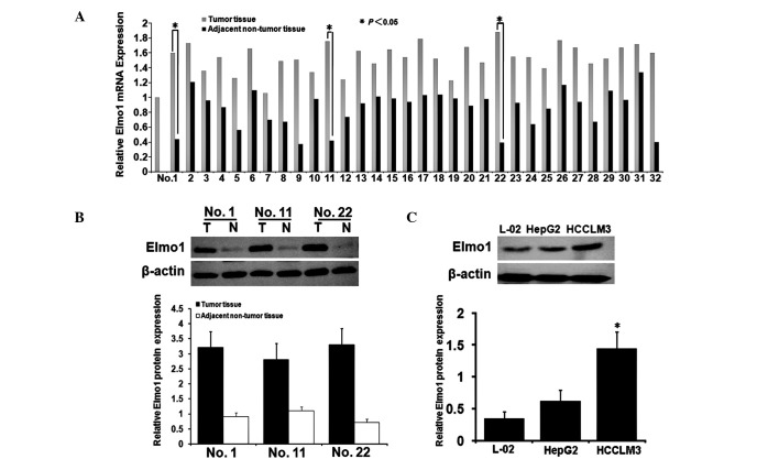
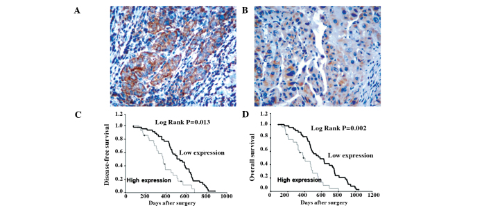
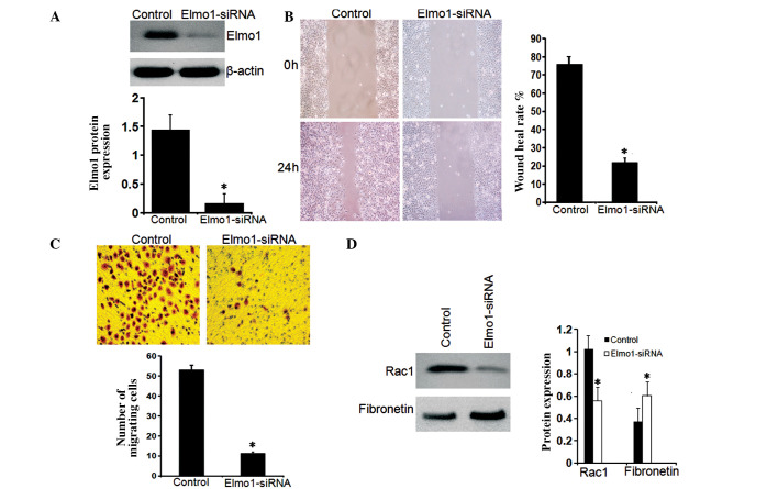
Engulfment and cell motility 1 (Elmo1) has been linked to the invasive phenotype of glioma cells. The use of Elmo1 inhibitors is currently being evaluated in hepato-cellular carcinoma (HCC), but the molecular mechanisms of their therapeutic effect have yet to be determined. Elmo1 expression in HCC tissue samples from 131 cases and in 5 HCC cell lines was determined by immunohistochemistry, quantitative RT-PCR and Western blotting. To functionally characterize Elmo1 in HCC, Elmo1 expression in the HCCLM3 cell line was blocked by siRNA. Cell migration was measured by wound healing and transwell migration assays in vitro. Elmo1 overexpression was significantly correlated with cell invasion and the poor prognosis of HCC. Elmo1-siRNA-treated HCCLM3 cells demonstrated a reduction in cell migration. The present study demonstrated for the first time that the suppression of Elmo1 expression inhibits cell invasion in HCC.


Figures

Figure 1.
Figure 1.
Expression levels of Elmo1 mRNA and protein in the HCC tissues and the adjacent non-tumor tissues (ANTTs). (A) Expression levels of Elmo1 mRNA in the 32 cases of HCC tissues and ANTTs. (B) Expression levels of Elmo1 protein in the HCC tissues (T) and ANTTs (N), respectively. Expression of Elmo1 mRNA and protein in the HCC tissues was significantly higher than that in the ANTTs (P<0.05). (C) Western blot analysis of Elmo1 protein expression was performed in the HepG2 and HCCLM3 HCC cell lines and in L-02 cells as a control. β-actin was used as the internal control for equal loading. The results indicated that the HCCLM3 cell line may be an ideal target for Elmo1 suppression.
Figure 2.
Figure 2.
Immunohistochemical detection of the protein expression of Elmo1 in HCC tissue and adjacent non-tumorous tissue (ANTT). (A) Positive staining for Elmo1 was noted in HCC tissue (original magnification, x400). (B) Weak staining for Elmo1 was noted in the pericarcinomatous liver tissue (original magnification, x400). Estimated (C) disease-free survival and (D) overall survival according to the expression of Elmo1 in 131 cases of HCC by the Kaplan-Meier method. Based on the results of immunohistochemical staining, the expression of Elmo1 was classified as low expression (0 or 1+; n=52) and high expression (2+ or 3+; n=79). The log-rank test shows that HCC patients with low Elmo1 expression had a longer median overall survival time and median disease-free survival time than patients with high expression.
Figure 3.
Figure 3.
Effect of gene silencing using Elmo1 siRNA on HCC cell migration and invasion. (A) After treatment of HCCLM3 cells with Elmo1 siRNA, Elmo1 protein expression in the normal and Elmo1-siRNA HCCLM3 cells was analyzed by SDS-PAGE and immunoblotting. β-actin expression levels were used to verify the equal loading of cellular protein. (B) After treatment with Elmo1 siRNA, a cell migration assay was performed. HCCLM3 cells (1×10⁶) were seeded on 6-cm plates coated with 10 μg/ml type I collagen. The cells were incubated for 24 h and the monolayer was disrupted with a cell scraper (1.2-mm diameter), then images were captured at 0 and 24 h under a phase contrast microscope. Experiments were carried out in triplicate, and four fields of each plate were assessed. (C) For the Matrigel invasion assay, HCCLM3 cells (2×10⁵) were seeded into the upper chamber of the Transwell, and the lower chamber was filled with 0.8 ml NIH-3T3 conditioned medium to induce chemotaxis. After 24 h of incubation at 37°C, the cells that invaded through the pores to the lower surface of the filter were counted under a microscope. Three invasion chambers were used per condition. The values obtained were calculated by averaging the total number of cells from three filters, and statistical analysis was performed using the Student's t-test. The data are expressed as the mean ± SD of at least three independent studies. (D) The reduced expression of Rac1 and increased expression of fibronectin were also noted in the Elmo1-siRNA HCCLM3 cells, indicating that Elmo1 expression may be involved in cell adhesion to the extracellular matrix and cell migration.

Similar articles

Cited by

References

    1. He J, Gu D, Wu X, Reynolds K, Duan X, Yao C, Wang J, Chen CS, Chen J, Wildman RP, Klag MJ, Whelton PK. Major causes of death among men and women in China. N Engl J Med. 2005;353:1124–1134. - PubMed
    1. Fan ST, Lo CM, Liu CL, Lam CM, Yuen WK, Yeung C, Wong J. Hepatectomy for hepatocellular carcinoma: toward zero hospital death. Ann Surg. 1999;229:322–330. - PMC - PubMed
    1. Hubert C, Sempoux C, Rahier J, Horsmans Y, Geubel A, van Beers BE, Annet L, Zech F, Leonard D, Gigot JF. Prognostic risk factors of survival after resection of hepatocellular carcinoma. Hepatogastroenterology. 2007;54:1791–1797. - PubMed
    1. Ibrahim S, Roychowdhury A, Hean TK. Risk factors for intrahepatic recurrence after hepatectomy for hepatocellular carcinoma. Am J Surg. 2007;194:17–22. - PubMed
    1. Mann CD, Neal CP, Garcea G, Manson MM, Dennison AR, Berry DP. Prognostic molecular markers in hepatocellular carcinoma: a systematic review. Eur J Cancer. 2007;43:979–992. - PubMed

LinkOut - more resources