. 2011 Nov;2(6):1047-1052.
doi: 10.3892/etm.2011.349. Epub 2011 Sep 5.

Anti-vascular endothelial growth factor antibody single therapy for pancreatic neuroendocrine carcinoma exhibits a marked tumor growth-inhibitory effect

Affiliations

Anti-vascular endothelial growth factor antibody single therapy for pancreatic neuroendocrine carcinoma exhibits a marked tumor growth-inhibitory effect

Kazuhiko Kasuya et al. Exp Ther Med. 2011 Nov.

Abstract

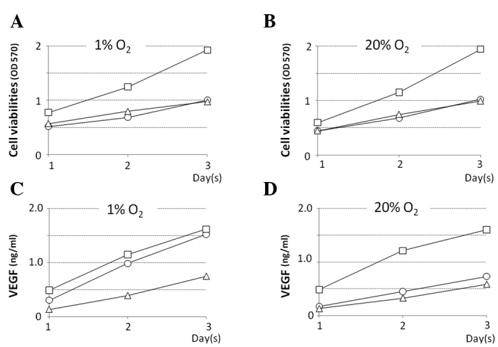
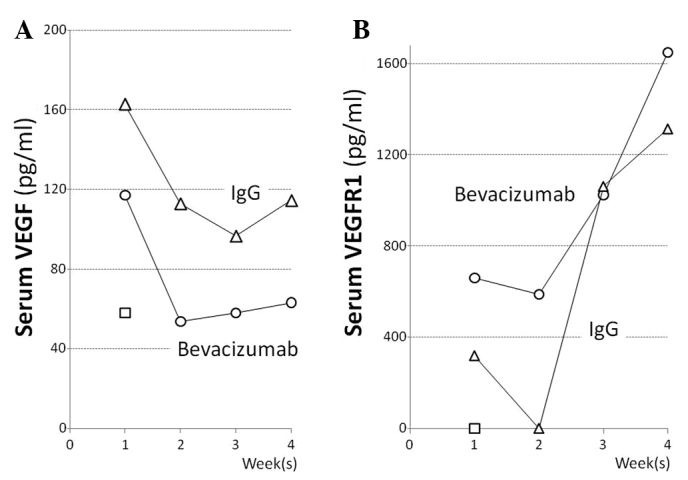
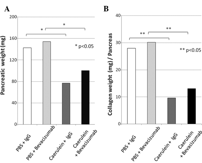
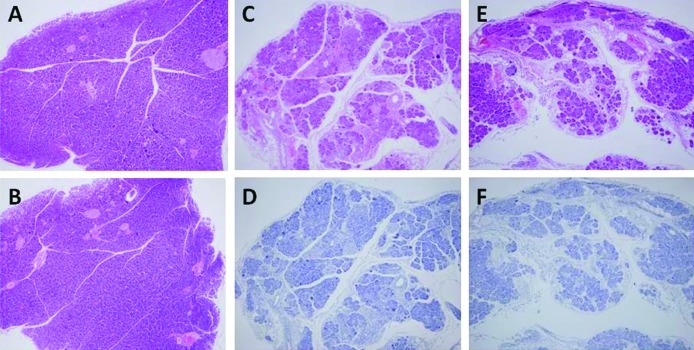
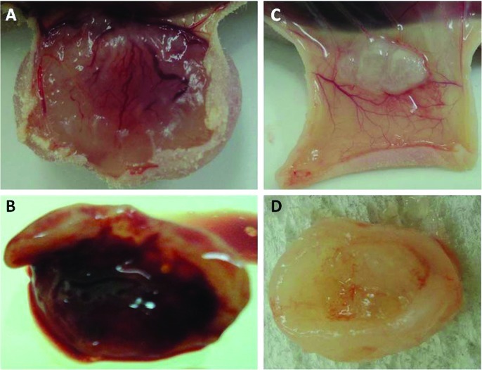
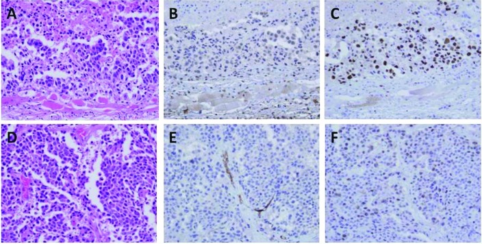
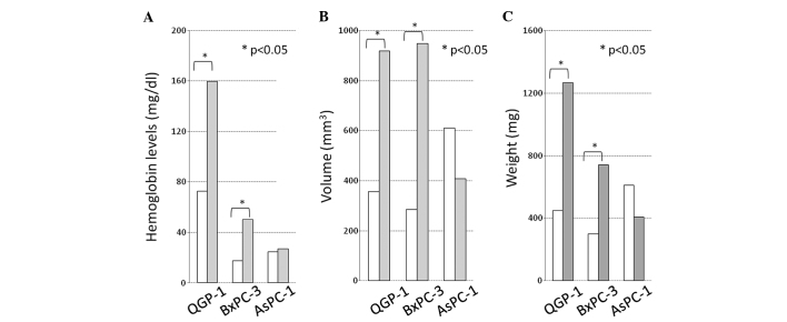
At present, no effective chemotherapy for pancreatic neuroendocrine carcinoma (PNEC) exists. However, anti-angiogenic therapy is expected to be effective for PNEC, a hypervascular tumor. We treated PNEC and hypovascular pancreatic ductal cell carcinoma (DCC) cell lines with the anti-vascular endothelial growth factor (VEGF) antibody bevacizumab, and compared the antitumor effect between the two different types of cell lines. The PNEC cell line QGP-1 and the DCC cell lines BxPC-3 and AsPC-1 were used. We evaluated the ability of the cell lines to proliferate and secrete VEGF in vitro, the antitumor effect of bevacizumab administration in vivo and the side effects of bevacizumab on the pancreas in a caerulein-induced pancreatitis model. Comparison of the QGP-1 and DCC cell lines showed that QGP-1 secreted a higher level of VEGF under a hypoxic environment than the DCC cell line, and bevacizumab exerted the most marked growth-inhibitory effect on QGP-1; the number of intratumoral blood vessels decreased and the percentage of proliferating cells was approximately the same. In the pancreatitis model, bevacizumab administration did not adversely affect the pancreatitis or the associated hypoxic environment. Bevacizumab does not affect the pancreas itself; therefore, its potent inhibitory effect on the growth of pancreatic neuroendocrine tumors alone can be expected.


Figures

Figure 1.
Figure 1.
Cell viability under a (A) 1% O2 and (B) 20% O2 environment. X-axis, time (in days). Y-axis, cell viability (absorbance). In all of the cell lines, the cell proliferation potency in an anoxia environment was the same as that in a normal oxygen environment. Level of VEGF (ng/ml) in the different culture mediums under a (C) 1% O2 and (D) 20% O2 environment. X-axis, time (in days). Y-axis, levels of VEGF (ng/mg). BxPc-3 (open squares), QGP-1 (open circles) and AsPC-1 (open triangles) cells. The VEGF secretional capacity of QGP-1 was increased in the anoxia environment.
Figure 2.
Figure 2.
Levels of (A) VEGF and (B) VEGFR1 in the serum of the mice. X-axis, time (in weeks). Y-axis, levels of VEGF (pg/mg) or VEGFR1 (pg/ml). Pancreatitis mice administered bevacizumab i.p. (open circles), mice administered human IgG i.p. as a control (open triangles) and normal mice (open squares).
Figure 3.
Figure 3.
(A) Pancreatic weight and (B) component density of collagen. The pancreatic weight and its component density of collagen significantly decreased in the pancreatitis mice. These changes due to bevacizumab administration were slight. IgG-administered mice (white bar), bevacizumab-administered normal mice (light grey bar), IgG-administered mice with caerulein-induced pancreatitis (dark grey bar) and bevacizumab-administered mice with pancreatitis (black bar).
Figure 4.
Figure 4.
Histological findings of the pancreas. (A) IgG- and (B) bevacizumab-administered normal mice. (C and D) IgG- and (E and F) bevacizumab-administered mice with caerulein-induced pancreatitis. (A, B, C and E) H&E staining; (D and F) pimonidasole staining.
Figure 5.
Figure 5.
Macroscopic view of subcutaneous tumors. Tumor and subcutaneous tissue of IgG-administered mice bearing (A) QGP-1 and (B) BxPC-3 cells. Tumor and subcutaneous tissue of bevacizumab-administered mice bearing (C) QGP-1 and (D) BxPC-3 cells.
Figure 6.
Figure 6.
Tumor and subcutaneous tissue in (A–C) bevacizumab- and (D–F) IgG-administered mice bearing QGP-1 cells. (A and B) H&E staining, (B and E) CD-34 staining and (C and F) Ki-67 staining. CD-34 immunoreactivity in the cancerous area in tumors of bevacizumab-administered mice was stronger than that of the IgG-administered mice.
Figure 7.
Figure 7.
(A) Hemoglobin levels, (B) volume (mm3) and (C) weight (mg) of the tumors. Tumors of bevacizumab-administered mice (white bar) and tumors of IgG-administered mice (grey bar). The hemoglobin levels, the volume and the weight of the tumors achieved significant differences between the bevacizumab- and IgG-administered mice bearing QGP-1 and BxPC-3 cells. *p<0.05.

References

    1. Eriksson B, Oberg K. Neuroendocrine tumours of the pancreas. Br J Surg. 2000;87:129–131. - PubMed
    1. Modlin IM, Oberg K, Chung DC, et al. Gastroenteropancreatic neuroendocrine tumours. Lancet Oncol. 2008;9:61–72. - PubMed
    1. Oberg KE, Reubi JC, Kwekkeboom DJ, Krenning EP. Role of somatostatins in gastroenteropancreatic neuroendocrine tumor development and therapy. Gastroenterology. 2010;139:742–753. - PubMed
    1. Kazanjian KK, Reber HA, Hines OJ. Resection of pancreatic neuroendocrine tumors: results of 70 cases. Arch Surg. 2006;141:765–769. - PubMed
    1. Takahashi Y, Akishima-Fukasawa Y, Kobayashi N, et al. Prognostic value of tumor architecture, tumor-associated vascular characteristics, and expression of angiogenic molecules in pancreatic endocrine tumors. Clin Cancer Res. 2007;13:187–196. - PubMed

LinkOut - more resources