. 2012 Aug;2(8):120104.
doi: 10.1098/rsob.120104.

Structured illumination of the interface between centriole and peri-centriolar material

Affiliations

Structured illumination of the interface between centriole and peri-centriolar material

Jingyan Fu et al. Open Biol. 2012 Aug.

Abstract

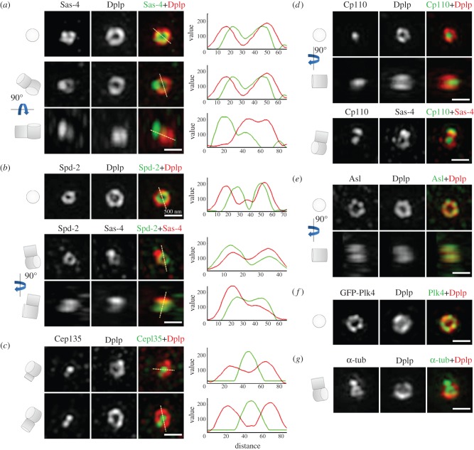
The increase in centrosome size in mitosis was described over a century ago, and yet it is poorly understood how centrioles, which lie at the core of centrosomes, organize the pericentriolar material (PCM) in this process. Now, structured illumination microscopy reveals in Drosophila that, before clouds of PCM appear, its proteins are closely associated with interphase centrioles in two tube-like layers: an inner layer occupied by centriolar microtubules, Sas-4, Spd-2 and Polo kinase; and an outer layer comprising Pericentrin-like protein (Dplp), Asterless (Asl) and Plk4 kinase. Centrosomin (Cnn) and γ-tubulin associate with this outer tube in G2 cells and, upon mitotic entry, Polo activity is required to recruit them together with Spd-2 into PCM clouds. Cnn is required for Spd-2 to expand into the PCM during this maturation process but can itself contribute to PCM independently of Spd-2. By contrast, the centrioles of spermatocytes elongate from a pre-existing proximal unit during the G2 preceding meiosis. Sas-4 is restricted to the microtubule-associated, inner cylinder and Dplp and Cnn to the outer cylinder of this proximal part. γ-Tubulin and Asl associate with the outer cylinder and Spd-2 with the inner cylinder throughout the entire G2 centriole. Although they occupy different spatial compartments on the G2 centriole, Cnn, Spd-2 and γ-tubulin become diminished at the centriole upon entry into meiosis to become part of PCM clouds.

Keywords: Drosophila; centriole; pericentriolar material; super resolution microscopy.


Figures

Figure 1.
Figure 1.
Localization of centriolar proteins in the centrosomes of cultured D.Mel-2 cells. D.Mel-2 cells stained to reveal the indicated centriolar proteins. (a) Sas-4 (green) and Dplp (red). The upper row shows a centriole before a procentriole becomes visible. Sas-4 is present in a similar cylindrical structure as Dplp but of a smaller diameter. As the daughter centriole elongates, it is loaded with Sas-4 (middle and bottom rows), whereas Dplp only decorates the mother centrioles. (b) Spd-2 (green) and Dplp (red), or Spd-2 (green) and Sas-4 (red). Spd-2 is present in a cylinder within the Dplp (upper row) and similar in diameter to the Sas-4 cylinder. Sas-4 is present on mother and daughter centrioles while Spd-2 predominantly on the mother centriole (middle and bottom rows). (c) Cep135 (green) and Dplp (red). Cep135 is present in the innermost part of mother centrioles and recruited to procentrioles in levels reflecting elongation. By contrast, Dplp preferentially associates with the mother centrioles, regardless of whether the daughter centrioles fully elongate. (d) Cp110 (green) and Dplp (red). Top view shows Cp110 to occupy the central most part of the centriolar cylinder (upper row). Side view shows Cp110 as a plug-like body at the distal end of a single centriole (middle row). Staining to reveal Cp110 (green) and Sas-4 (red) shows that Cp110 is present at the distal end of both mother and daughter centrioles (bottom row). (e) Asl (green) and Dplp (red) showing their overlapping distribution in cylindrical structures. Asl shows a punctate staining pattern within the nine-fold symmetrical structure. (f) D.Mel-2 cells expressing GFP-Plk4 (green) and stained to reveal Dplp (red). The distribution of Plk4 strongly resembles that of its partner protein Asl. (g) D.Mel-2 cells stained to reveal α-tubulin (α-tub; green) and Dplp (red). α-tubulin is present in a cylinder that lies within the region of Dplp staining and that presumably corresponds to the nine sets of microtubules. It is also seen in the daughter centriole. Scale bars, (ag) 500 nm.
Figure 2.
Figure 2.
Scheme depicting the structure of a centrosome and the distribution of different proteins. (a,b) The mother centriole is shown partitioned into five zones corresponding to the zones of staining that we described. Zones III, IV and part of the zone II (proteins without asterisk) are absent from the daughter centriole. Also, parts of the zone III (proteins marked with #) only start to accumulate in late G2 phase. The proteins present in each zone are listed in the table, and the diameters (±s.d.) of the zones are defined by the staining patterns as indicated. tub, tubulin.
Figure 3.
Figure 3.
Development of the pericentriolar material (PCM) during centrosome maturation. (ac) D.Mel-2 cells were stained to reveal (a) Spd-2 (green) and Dplp (red), (b) Cnn (green) and Dplp (red), or (c) γ-tubulin (green) and Dplp (red). Representative interphase and metaphase centrioles are shown. Spd-2 localizes to all interphase centrioles, whereas Cnn and γ-tubulin are found only on a small subset of interphase centrioles. All three proteins display cylindrical structures in interphase with the diameters of Cnn and γ-tubulin cylinders being similar to those of Dplp. Spd-2, on the other hand, is more compact around the centriole core. During mitosis, there is a large accumulation of pericentriolar Spd-2, Cnn and γ-tubulin with only Spd-2 being still detectable at the core of the centriole itself. Scale bars: 500 nm. (d) D.Mel-2 cells expressing GFP-Polo were stained to reveal Dplp. Centrosomes from interphase (I) and metaphase (II) cells are shown. In interphase cells, Polo is present both in centrioles as a cylinder within the Dplp cylinder and also on cytoplasmic microtubules. In metaphase, Polo extends into the PCM but not as extensively as Spd-2, Cnn and γ-tubulin. Scale bar in upper row: 4 μm; lower rows: 500 nm.
Figure 4.
Figure 4.
Interdependence of Polo, Cnn and Spd-2 in centrosome maturation. (a,b) D.Mel-2 cells were treated with dimethylsulfoxide (DMSO) or BI2536 for 16 h, or with glutathione S-transferase (GST), Spd-2 or Cnn dsRNA, and stained to reveal γ-tubulin (green), Dplp (red) and phospho-Histone H3 Ser10 (not shown). Note that in following inhibition of Polo or depletion of Spd-2 or Cnn, γ-tubulin is removed from PCM and only a remnant remains on the centriole. Quantitation of fluorescence intensity of γ-tubulin is normalized to that of Dplp. Error bars represent s.e.m. and asterisks indicate p < 0.0001. (c,d) D.Mel-2 cells were treated with DMSO or BI2536 for 16 h, or with GST or Cnn dsRNA, and stained to reveal Spd-2 (green), Dplp (red) and phospho-Histone H3 Ser10 (not shown). Note that both Polo inhibition and Cnn depletion completely remove Spd-2 from PCM, but the inner cylindrical structure of Spd-2 in the centriole remains intact. Quantitation of total fluorescence intensity of Spd-2 in the pericentriolar region is normalized to that of Dplp. This required subtracting fluorescence of inner cylindrical structures. Error bars represent s.e.m. and asterisks indicate p < 0.0001. (e,f) D.Mel-2 cells were treated with DMSO or BI2536 for 16 h, or with GST or Spd-2 dsRNA and stained to reveal Cnn (green), Dplp (red) and phospho-Histone H3 Ser10 (not shown). Quantitation shows fluorescence intensity of Cnn or total volume occupied by Cnn normalized to that of Dplp. Error bars represent s.e.m. and asterisks indicate p < 0.0001. Whereas Cnn is completely removed from the PCM after BI2536 treatment, after Spd-2 depletion the fluorescence intensity of Cnn is reduced by 56% and the volume it occupies by 49%. (g) Schematic showing the hierarchy of PCM assembly. Polo localizes to the centriole and extends only a little into the PCM. Spd-2 occupies a similar part of the centriole but is more diffuse in PCM. Cnn and γ-tubulin, on the other hand, are harboured only in PCM, lying outwards of Dplp. During PCM assembly, Polo, that could be either cytoplasmic or centriole-associated, activates Cnn, allowing it to localize to PCM. Both of them are subsequently required for Spd-2 recruitment to the PCM. Cnn cooperates with Spd-2 to recruit γ-tubulin. In addition, Spd-2 is also required to maintain Cnn levels. Scale bars, (a,c,e) 500 nm.
Figure 5.
Figure 5.
Growth of spermatocyte centrioles in the extended G2 preceding meiosis. (a) Centrioles from cysts of primary spermatocytes at progressively later stages of G2 expressing GFP-PACT (green) were stained to reveal Dplp (red). Dplp remains associated with the hinge area and the proximal part of the centriole that increases in length from approximately 360 to 540 nm (red). During the same interval, centriolar microtubules marked with GFP-PACT increase in length from 250 nm to 1.4 μm (green). (b) Mature primary spermatocyte centrioles (still in G2) expressing GFP-PACT (green) and stained to reveal Cep135 (red). Note that Cep135 extends down the length of the GFP-PACT cylinder. (c) Mature primary spermatocyte centrioles stained to reveal Asl (green) and Dplp (red). Asl extends along the length of the centriole in a cylinder of similar diameter to Dplp, which is restricted to the proximal part; Sas-4 (green) and Dplp (red). Both Sas-4 and Dplp are restricted to the proximal part, with Sas-4 staining being contained within that of Dplp. (d) Immature primary spermatocyte centrioles expressing GFP-PACT or GFP-Cep135 (green) were stained to reveal Cp110 (red). Note that Cp110 shows localization at the distal ends separated from GFP-PACT. On the contrary, Cep135, which exceeds the distal end of PACT signal, shows spatial interaction with Cp110. Scale bars, (ad) 1 µm.
Figure 6.
Figure 6.
Localization of PCM proteins during development of primary spermatocytes. (ac) Spermatocyte centrioles in early and late G2 and in meiosis I and meiosis II stained to reveal Spd-2, Cnn, and γ-tubulin (green) and Dplp (red). In early and late G2 spermatocytes, Spd-2 localizes inside the Dplp tube but extends to the distal part. Cnn and γ-tubulin overlap with Dplp; γ-tubulin also extends along the length of the centriole, while Cnn is predominantly but not exclusively at the proximal end. During meiosis I and II, Spd-2, Cnn and γ-tubulin all expand into the PCM; Cnn is more closely associated with the centriole core than Spd-2 and γ-tubulin. Scale bars, (ac) 1 µm.

References

    1. Wilson EB. 1896; 1900; 1925. The cell in development and inheritance. New York, NY: Macmillan
    1. Boveri T. 1914. Zur Frage der Entstehung maligner Tumoren. Jena, Germany: Gustav Fischer Verlag
    1. Boveri T. 1888. Zellenstudien II. Die Befruchtung und Teilung des Eies von Ascaris megalocephala. Jena Zeit. Naturw. 22, 685–882
    1. Strnad P, Gonczy P. 2008. Mechanisms of procentriole formation. Trends Cell Biol. 18, 389–39610.1016/j.tcb.2008.06.004 (doi:10.1016/j.tcb.2008.06.004) - DOI - DOI - PubMed
    1. Mahen R, Venkitaraman AR. 2012. Pattern formation in centrosome assembly. Curr. Opin. Cell Biol. 24, 14–2310.1016/j.ceb.2011.12.012 (doi:10.1016/j.ceb.2011.12.012) - DOI - DOI - PubMed

Publication types

MeSH terms

LinkOut - more resources