

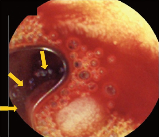
Ectopic pancreas bleeding in the jejunum revealed by capsule endoscopy
- PMID: 22977800
- PMCID: PMC3429734
- DOI: 10.5946/ce.2012.45.3.194
Ectopic pancreas bleeding in the jejunum revealed by capsule endoscopy
Abstract
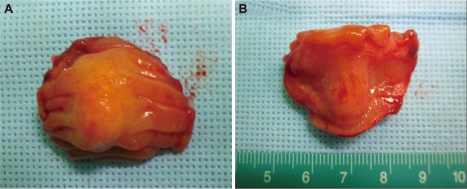
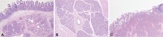
Ectopic pancreas is defined as pancreatic tissue found outside the usual anatomic location. It is often found incidentally at different sites in the gastrointestinal (GI) tract. The incidence of ectopic pancreatic tissue in autopsy series is 1% to 2%, with 70% of the ectopic lesions found in the stomach, duodenum and jejunum. Although it is usually a silent anomaly, an ectopic pancreas may become clinically evident when complicated by inflammation, bleeding, obstruction or malignant transformation. We report a case of ectopic pancreas located in the jejunum and presenting as an obscure GI bleeding, which was diagnosed by capsule endoscopy.
Keywords: Capsule endoscopy; Ectopic pancreas; Gastrointestinal hemorrhage.
Conflict of interest statement
The authors have no financial conflicts of interest.
Figures
References
-
- Lai EC, Tompkins RK. Heterotopic pancreas. Review of a 26 year experience. Am J Surg. 1986;151:697–700. - PubMed
-
- Armstrong CP, King PM, Dixon JM, Macleod IB. The clinical significance of heterotopic pancreas in the gastrointestinal tract. Br J Surg. 1981;68:384–387. - PubMed
-
- Chen HL, Chang WH, Shih SC, Bair MJ, Lin SC. Changing pattern of ectopic pancreas: 22 years of experience in a medical center. J Formos Med Assoc. 2008;107:932–936. - PubMed
-
- Dolan RV, ReMine WH, Dockerty MB. The fate of heterotopic pancreatic tissue. A study of 212 cases. Arch Surg. 1974;109:762–765. - PubMed
-
- Chen HL, Lin SC, Chang WH, Yang TL, Chen YJ. Identification of ectopic pancreas in the ileum by capsule endoscopy. J Formos Med Assoc. 2007;106:240–243. - PubMed
LinkOut - more resources
Full Text Sources
