

Endoscopic resection of subepithelial tumors
- PMID: 22977810
- PMCID: PMC3429744
- DOI: 10.5946/ce.2012.45.3.240
Endoscopic resection of subepithelial tumors
Abstract
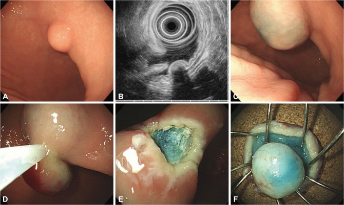
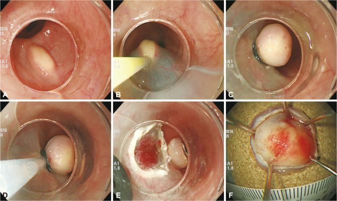
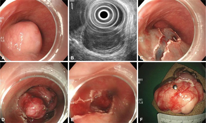
Subepithelial tumors (SETs) are often incidentally found during endoscopic examinations. Endoscopic ultrasonography (EUS) is a good method for differential diagnosis of SETs, but a definite diagnosis cannot be made based on EUS features alone in some cases. Periodic follow-up examinations by endoscopy and EUS remains the recommended management strategy, which involves issues related to patient compliance, cost-effectiveness, and the risk associated with repeated endoscopic procedures and delayed diagnosis of malignancy. Endoscopic resection of the SETs is another technique to treat them as well as to obtain tissue specimens for accurate histologic diagnosis. Herein, a various endoscopic techniques ranging from simple snare resection to endoscopic submucosal tunnel dissection for the management of SETs will be reviewed.
Keywords: Endoscopic resection; Neoplasms; Subepithelial.
Conflict of interest statement
The author has no financial conflicts of interest.
Figures
References
-
- Hedenbro JL, Ekelund M, Wetterberg P. Endoscopic diagnosis of submucosal gastric lesions. The results after routine endoscopy. Surg Endosc. 1991;5:20–23. - PubMed
-
- Nickl N. Endoscopic approach to gastrointestinal stromal tumors. Gastrointest Endosc Clin N Am. 2005;15:455–466. - PubMed
-
- Chak A. EUS in submucosal tumors. Gastrointest Endosc. 2002;56(4 Suppl):S43–S48. - PubMed
-
- Hwang JH, Rulyak SD, Kimmey MB American Gastroenterological Association Institute. American Gastroenterological Association Institute technical review on the management of gastric subepithelial masses. Gastroenterology. 2006;130:2217–2228. - PubMed
-
- Hwang JH, Kimmey MB. The incidental upper gastrointestinal subepithelial mass. Gastroenterology. 2004;126:301–307. - PubMed
LinkOut - more resources
Full Text Sources
