Review
. 2013 Sep 1;19(7):708-22.
doi: 10.1089/ars.2012.4932. Epub 2012 Oct 25.

Mono- and dithiol glutaredoxins in the trypanothione-based redox metabolism of pathogenic trypanosomes

Affiliations
Review

Mono- and dithiol glutaredoxins in the trypanothione-based redox metabolism of pathogenic trypanosomes

Marcelo A Comini et al. Antioxid Redox Signal. .

Abstract

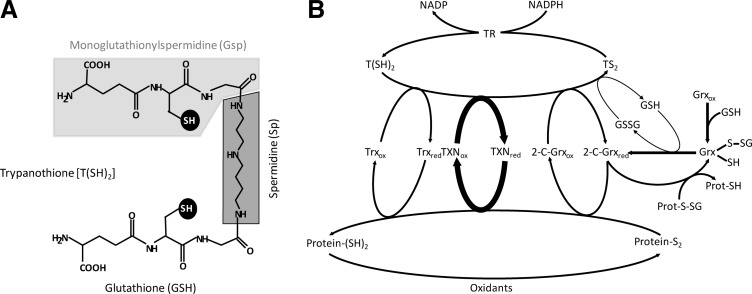
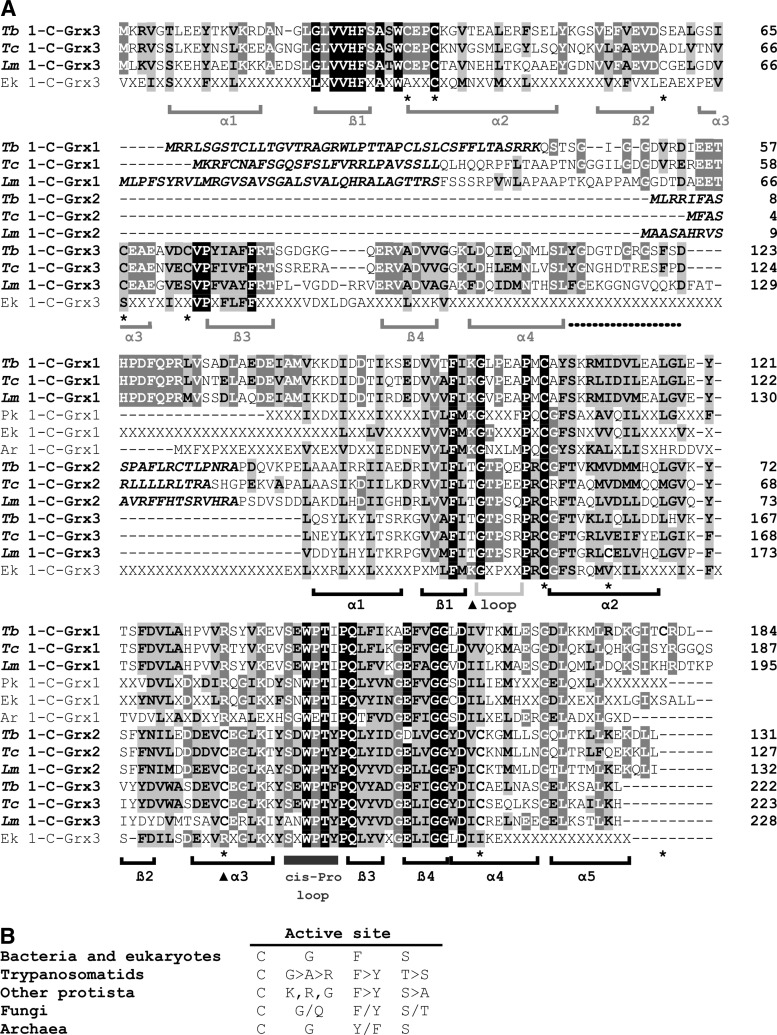
Significance: Glutaredoxins are ubiquitous small thiol proteins of the thioredoxin-fold superfamily. Two major groups are distinguished based on their active sites: the dithiol (2-C-Grxs) and the monothiol (1-C-Grxs) glutaredoxins with a CXXC and a CXXS active site motif, respectively. Glutaredoxins are involved in cellular redox and/or iron sulfur metabolism. Usually their functions are closely linked to the glutathione system. Trypanosomatids, the causative agents of several tropical diseases, rely on trypanothione as principal low molecular mass thiol, and their glutaredoxins readily react with the unique bis(glutathionyl) spermidine conjugate.

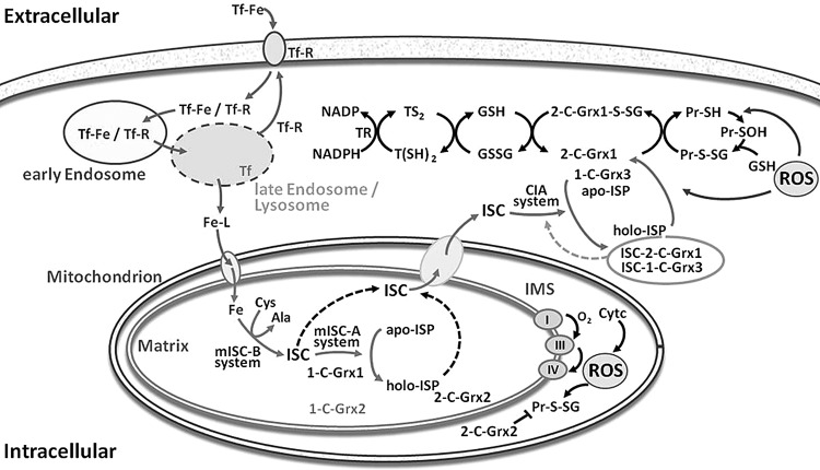
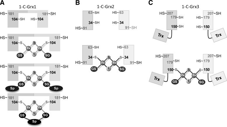
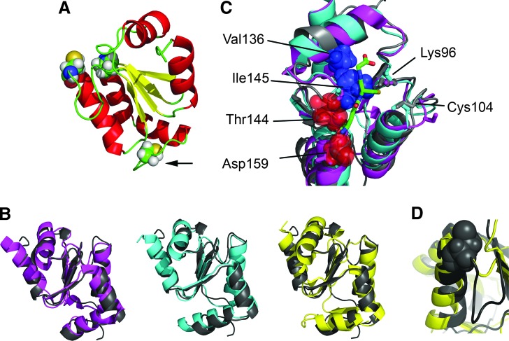
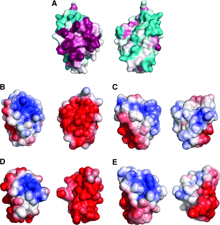
Recent advances: Two 2-C-Grxs and three 1-C-Grxs have been identified in pathogenic trypanosomatids. The 2-C-Grxs catalyze the reduction of glutathione disulfide by trypanothione and display reductase activity towards protein disulfides, as well as protein-glutathione mixed disulfides. In vitro, all three 1-C-Grxs as well as the cytosolic 2-C-Grx of Trypanosoma brucei can complex an iron-sulfur cluster. Recently the structure of the 1-C-Grx1 has been solved by NMR spectroscopy. The structure is very similar to those of other 1-C-Grxs, with some differences in the loop containing the conserved cis-Pro and the surface charge distribution.

Critical issues: Although four of the five trypanosomal glutaredoxins proved to coordinate an iron-sulfur cluster in vitro, the physiological role of the mitochondrial and cytosolic proteins, respectively, has only started to be unraveled.

Future directions: The use of trypanothione by the glutaredoxins has established a novel role for this parasite-specific dithiol. Future work should reveal if these differences can be exploited for the development of novel antiparasitic drugs.


Figures

FIG. 1.
FIG. 1.
The trypanothione-based redox metabolism of trypanosomatids. (A) The main low molecular mass thiol is trypanothione [T(SH)2]. It is synthesized from two molecules of glutathione (GSH, light gray background) and one spermidine (Sp, dark gray background) with mono(glutathionyl)spermidine (Gsp; gray backgrounds) as intermediate. The two thiol groups acting as redox cofactors or ligands are highlighted on a black background. (B) The NADPH-dependent flavoenzyme trypanothione reductase (TR) catalyzes the reduction of trypanothione disulfide (TS2) to T(SH)2 and, thus, is responsible for maintaining the cellular thiol redox homeostasis. T(SH)2 is the direct reducing agent for the parasite thioredoxin (Trx), tryparedoxin (TXN), and dithiol glutaredoxin (2-C-Grx) type oxidoreductases, as well as for glutathione disulfide (GSSG). In vitro all three oxidoreductases catalyze the reduction of intra and/or intermolecular protein disulfides (Protein-S2) to the respective reduced forms (Protein-(SH)2); however TXN is by far the most efficient multipurpose oxidoreductase of the parasites. Trx displays an extremely low cellular concentration and its physiological role, if any, is not known. The parasite Grxs also catalyze the reduction of protein disulfides and specifically, of glutathione–protein mixed disulfides (Prot-S-SG), as well as of GSSG by T(SH)2. The subindex “red” or “ox” denotes the dithiol (reduced) and disulfide (oxidized) form of the proteins.
FIG. 2.
FIG. 2.
Sequence analysis of monothiol glutaredoxins. (A) 1-C-Grxs from trypanosomatids (Tb, Trypanosoma brucei; Tc, Trypanosoma cruzi; Lm, Leishmania major) were aligned with the consensus sequences obtained for prokaryote (Pk), eukaryote (Ek), and archeabacteria (Ar) homologues using the ClustalW algorithm (85) and manually adjusted as necessary. Accession numbers are AJ619696, AM489503 and AM489504 for T. brucei 1-C-Grx1, 2 and 3, respectively; XP_807837, XP_803206 and XP_813048 for T. cruzi 1-C-Grx1, 2 and 3, respectively; NP_047037, CAJ01951 and XP_843232 for L. major 1-C-Grx1, 2 and 3, respectively. Residues given in italic indicate the predicted mitochondrial targeting sequence of Kinetoplastida 1-C-Grx1 and 2. Residues shown in black on gray and white on gray represent residues that are similar and identical, respectively, in at least 40% of the aligned sequences. Residues that are strictly conserved in all sequences analyzed are shown white on black. Cysteine residues in the 1-C-Grxs from trypanosomatids are indicated with an asterisk at the bottom of the alignment. The arrow heads mark basic residues suggested to be involved in glutathione binding in classical Grxs (28, 40). The consensus sequences for the three phylogenetic domains were obtained by alignment of characterized or putative 1-C-Grxs from representative organisms. The sequences used for prokaryotes were from Agrobacterium tumefaciens (α-protobacterium, Acc. Nr. AAK87621.1), Neisseria gonorrheae (β-protobacterium, YP_207507.1), Myxococcus xanthus (δ-protobacterium, ABF89434.1), Escherichia coli (γ-protobacterium, 1YKA) and Synechococcus elongatus (cyanobacterium, BAD78595.1); for archaeabacteria: Haloarcula marismortui (AAV46243.1), Natronomonas pharaonis (YP_326686.1), Haloquadratum walsbyi (CAJ51793.1), and Halobacterium sp. (AAG18993.1); for eukaryotes: Gallus gallus (NP_001008472.1 and XP_421826.1), Danio rerio (AAH59659.1 and NP_001005950.1), Homo sapiens (NP_057501.2 and AAH05289.1), Caenorhabditis elegans (CAB11547.1 and NP_001023756.1), Apis mellifera (XP_625213.1 and XP_392870.1), Tetraodon negroviridis (CAG00128.1 and CAG02746.1), Saccharomyces cerevisiae (Q02784, Q03835 and P32642), Arabidopsis thaliana (AY157988), Tribolium castaneum (XP_975383.1 and XP_972466.1), Porphyra purpurea (P51384), Bos taurus (XP_582303.1 and AAX46537.1), and Xenopus tropicalis (AAH75374.1 and NP_001017209.1). The consensus sequences show only residues that were common to more than 75% of the proteins analyzed. X represents any amino acid. The secondary structure motifs below the alignment refer to the α-helical (α) and ß-sheet (ß) regions in the NMR structure of T. brucei 1-C-Grx1 (black) as well as the homology model of the Trx domain of T. brucei 1-C-Grx3 (gray). The dotted line marks the linker region of T. brucei 1-C-Grx3 and the light and dark gray bar indicates the insertion preceding the active site and the cys-Pro loop. (B) The consensus active site motif of 1-C-Grxs from representatives of the prokaryotic and eukaryotic domains and the family of Trypanosomatidae was obtained from the sequences listed above. The following sequences were used for other protista: Cryptosporidium parvum (CAD98438), Tetrahymena thermophila (XP 001016225, XP 001032143 and XP 001008985), Paramecium tetraurelia (CAK57552, CAK90692 and CAK55785), and Plasmodium falciparum (CAG25239 and CAD50844, for Glp2 and Glp3, respectively); fungi: Encephalitozoon cuniculi (NP 597481), Cocidioides immitis (XP 001244791), and Mortierella alpina (CAB 56513); Archaea: Haloarcula morismortui (AAV46243), Natromonas pharaonis (YP326686), and Haloquadratum walsbyi (CAJ 51793).
FIG. 3.
FIG. 3.
Putative roles of the glutaredoxins in the thiol and iron metabolism of African trypanosomes. African trypanosomes obtain iron by the uptake of iron-loaded transferrin (Tf-Fe) from the host blood via receptor-mediated (Tf-R) endocytosis. In the late endosome, Fe is released and exported into the cytosol. Here it circulates in complex with low molecular mass ligands (Fe–L) and can be translocated into the mitochondrial matrix. As shown for yeast and mammalian cells as well as partially for trypanosomes, the mitochondrion is the primary site for the biosynthesis of ISC that requires two specialized machineries. The mitochondrial Iron Sulfur Cluster Biogenesis (mISC-B) system synthesizes the ISC from iron and cysteine (as sulfur donor), and the Iron Sulfur Cluster Assembly (mISC-A) system then transfers the pre-formed ISC from scaffold proteins to acceptor iron sulfur proteins (ISP). T. brucei 1-C-Grx1 is an indispensable (iron sulfur) protein, probably participating in the mISC-A system. The parasite 1-C-Grx2 is a low abundant and functionally nonredundant orthologue of 1-C-Grx1. How ISC is exported from the mitochondrion is not yet known. In the cytosol, 1-C-Grx3 and 2-C-Grx1 may be part of the cytosolic iron assembly (CIA) machinery that transforms apo-iron sulfur proteins (apo-ISP) into the respective holo-proteins (holo-ISP). Alternatively, ISC formation on 2-C-Grx1 may result in an inactive form that, in the presence of high levels of reactive oxygen species (ROS), may be converted again in the free protein (for further abbreviations, see legend of Fig. 1). 2-C-Grx2 in the—intermembrane space (IMS) and/or matrix of the—mitochondrion plays probably a crucial role for the reduction of protein disulfides and/or glutathionylated proteins (Pr-S-SG) produced by reactive oxygen species (ROS) originating from the respiratory chain and cytochrome c (cyt c) activity present only in the insect stage of the parasite. The scheme is based on data from reference 55 and citations therein.
FIG. 4.
FIG. 4.
Working models for iron–sulfur cluster assembly into 1-C-Grxs from trypanosomatids. (A) 1-C-Grx1 is a homodimeric protein in free form as well as after binding an ISC complex. ISC coordination involves the active site Cys104 (bold) from each subunit and two additional sulfhydryl groups provided by either two molecules of GSH (-SG) or Gsp (–SG-Sp) or by one trypanothione [(-SG)2-Sp] molecule. (B) and (C) Apo 1-C-Grx2 and 1-C-Grx3 are monomeric proteins that dimerize upon binding of an ISC. Depicted are the putative complexes involving the active site Cys34 and Cys150 (bold), respectively, and two molecules of GSH as nonprotein ligands.
FIG. 5.
FIG. 5.
Three-dimensional structure of T. brucei 1-C-Grx1 in comparison with other 1-C-Grxs. (A) The structure of T. brucei 1-C-Grx1 (residues 76–184) was determined by multidimensional NMR spectroscopy (PDB ID 2LTK). The active site Cys104, the conserved cis-Pro146 facing the active site (upper left part) and the only other cysteine residue (Cys181, marked by an arrow) are shown as spheres. (B) Overlay of the solution structure of T. brucei 1-C-Grx1 (gray) with the crystal structures of 1-C-Grxs: S. cerevisiae Grx5 (magenta, PDB-id 3GX8, backbone-RMSD: 1.5 Å), E. coli Grx4 (cyan, PDB-id 2WCI, backbone-RMSD: 1.5 Å) and S. cerevisiae Grx6 (yellow, PDB-id 3L4N, backbone-RMSD: 1.7 Å). The abbreviations for the proteins correspond to those in the respective publication. (C) Superposition of the structures of T. brucei 1-C-Grx1 (gray; PDB-ID 2LTK), E. coli Grx4 in ISC-bound dimeric form (cyan; PDB-ID 2WIC; a single monomer is shown with the noncovalently bound GSH; the ISC is omitted), and S. cerevisiae Grx5 (magenta, PDB-ID 3GX8). Residues shown to be critical for GSH binding and ISC ligation are depicted as sticks (corresponding to Lys96, Cys104, Thr144, and Asp159 in T. brucei 1-C-Grx); the side chains of Val136 and Thr144, Ile145, and Asp159 in T. brucei 1-C-Grx1 are indicated with spheres; the glutathione moiety from the structure of E. coli holo-Grx4 is shown in green. It is evident that the change in the conformation of the loop containing the cis-Pro146 observed in T. brucei 1-C-Grx1 significantly distorts the GSH pocket and precludes the binding of glutathione as determined in E. coli Grx4. (D) Enlargement of the active site region of T. brucei 1-C-Grx1 (gray) and S. cerevisiae Grx6 (yellow) showing the presence and absence of the loop that precedes the active site in the corresponding protein. To see this illustration in color, the reader is referred to the web version of this article at www.liebertpub.com/ars
FIG. 6.
FIG. 6.
Molecular surface of T. brucei 1-C-Grx1 and 3. (A) Surface amino acid conservation in T. brucei 1-C-Grx1 calculated with the Consurf server using the 150 unique 1-C-Grx sequences with the lowest E-value from PSI-BLAST. Conserved and variable residues are depicted in purple and cyan, respectively. Electrostatic potential mapped onto the molecular surface of (B) T. brucei 1-C-Grx1, (C) T. brucei C-terminal domain of Grx3, (D) S. cerevisiae Grx5, and (E) E. coli Grx4; red and blue denote negatively and positively charged residues, respectively. Each image shows two views of the same protein rotated by z-180°. To see this illustration in color, the reader is referred to the web version of this article at www.liebertpub.com/ars

Similar articles

Cited by

References

    1. Alphey MS. Gabrielsen M. Micossi E. Leonard GA. McSweeney SM. Ravelli RB. Tetaud E. Fairlamb AH. Bond CS. Hunter WN. Tryparedoxins from Crithidia fasciculata and Trypanosoma brucei: photoreduction of the redox disulfide using synchrotron radiation and evidence for a conformational switch implicated in function. J Biol Chem. 2003;278:25919–25925. - PubMed
    1. Arias DG. Cabeza MS. Erben ED. Carranza PG. Lujan HD. Tellez Inon MT. Iglesias AA. Guerrero SA. Functional characterization of methionine sulfoxide reductase A from Trypanosoma spp. Free Radic Biol Med. 2011;50:37–46. - PubMed
    1. Askelöf P. Axelsson K. Eriksson S. Mannervik B. Mechanism of action of enzymes catalyzing thiol-disulfide interchange. Thioltransferases rather than transhydrogenases. FEBS Lett. 1974;38:263–267. - PubMed
    1. Atkinson H. Babbitt PC. An atlas of the thioredoxin fold class reveals the complexity of function-enabling adaptations. PLoS Comput. Biol. 2009;5:e1000541. - PMC - PubMed
    1. Beer SM. Taylor ER. Brown SE. Dahm CC. Costa NJ. Runswick MJ. Murphy MP. Glutaredoxin 2 catalyzes the reversible oxidation and glutathionylation of mitochondrial membrane thiol proteins: Implications for mitochondrial redox regulation and antioxidant defense. J Biol Chem. 2004;279:47939–47951. - PubMed

Publication types

LinkOut - more resources