

Normalization of hindbrain morphology after decompression of Chiari malformation Type I
- PMID: 22978540
- PMCID: PMC3786329
- DOI: 10.3171/2012.8.JNS111476
Normalization of hindbrain morphology after decompression of Chiari malformation Type I
Abstract
Object: Chiari malformation Type I (CM-I) is characterized by hindbrain deformity. We investigated the effects of craniocervical decompression surgery on the anatomical features of hindbrain deformity with a prospective MRI study of patients with CM-I.
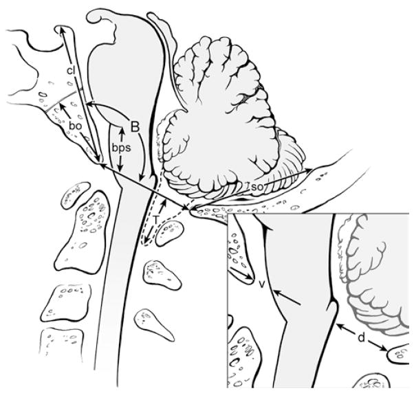
Methods: A prospective longitudinal study was conducted in 48 patients with CM-I (39 with syringomyelia) treated with craniocervical decompression. Clinical examinations and cervical MRI were performed before surgery and 1 week, 3-6 months, and annually after surgery. Hindbrain deformity was defined by tonsillar ectopia, pointed cerebellar tonsils, and/or cervicomedullary protuberance. The length of the clivus, basiocciput (sphenooccipital synchondrosis to basion), supraocciput (internal occipital protuberance to opisthion), and anteroposterior (AP) width of CSF pathways at the foramen magnum were measured and compared with those from 18 healthy volunteers (control group).
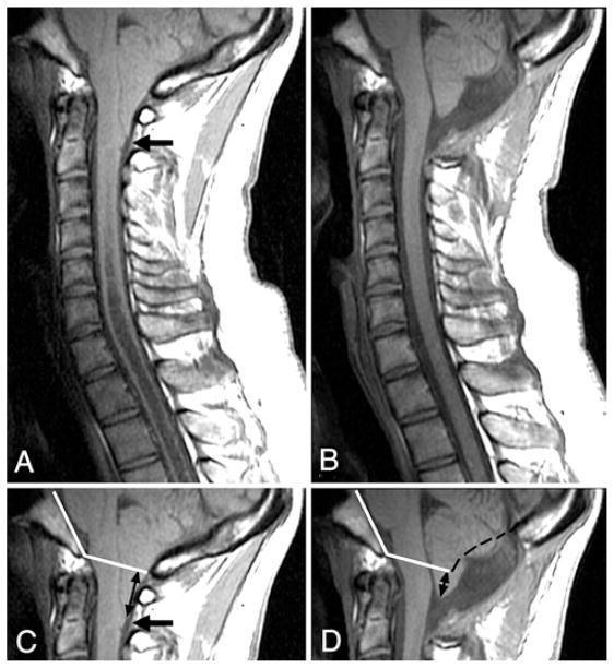
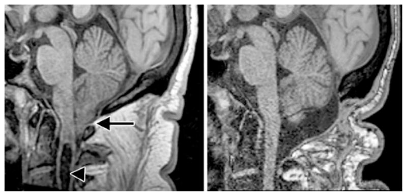
Results: Before surgery, the patients' posterior fossa bones were short and their CSF pathways were narrow. All patients had tonsillar ectopia (mean [± SD] 12.3 ± 5.1 mm; normal 0.3 ± 1.0). The majority of patients had pointed tonsils and more than two-thirds exhibited a cervicomedullary protuberance. Clivus and basiocciput lengths were significantly shorter than the values obtained in the control group. However, the supraocciput length did not differ significantly from control measurements. The mean bulbopontine sulcus distance superior to the basion was 9.5 ± 2.6 mm (vs 13.6 ± 2.8 mm in controls; p < 0.0001). The AP widths of the CSF pathways at the level of the foramen magnum were significantly narrowed. After surgery, CSF pathways significantly expanded both ventrally and dorsally. By 3-6 months after surgery, pointed tonsils became round, cervicomedullary protuberance disappeared, and tonsillar ectopia diminished by 51% (to 6.0 ± 3.3 mm; p < 0.0001).
Conclusions: The cerebellar tonsils and brainstem assumed a normal appearance within 6 months after craniocervical decompression. These findings support the concept that the CM-I is not a congenital malformation of the neural elements but rather an acquired malformation that arises from pulsatile impaction of the cerebellar tonsils into the foramen magnum. Clinical trial registration no.: NCT00001327.
Conflict of interest statement
The authors report no conflict of interest concerning the materials or methods used in this study or the findings specified in this paper.
Author contributions to the study and manuscript preparation include the following. Conception and design: Heiss, Oldfield. Acquisition of data: Heiss, Suffredini. Analysis and interpretation of data: all authors. Drafting the article: Heiss, Suffredini. Critically revising the article: all authors. Reviewed submitted version of manuscript: all authors. Approved the final version of the manuscript on behalf of all authors: Heiss. Statistical analysis: Suffredini, Bakhtian. Administrative/technical/material support: Bakhtian, Oldfield. Study supervision: Heiss, Oldfield.
Figures
References
-
- Chiari H. Concerning changes in the cerebellum due to hydrocephalus of the cerebrum. Dtsch Med Wochenschr. 1891;17:1172–1175. (Ger)
-
- Duddy MJ, Williams B. Hindbrain migration after decompression for hindbrain hernia: a quantitative assessment using MRI. Br J Neurosurg. 1991;5:141–152. - PubMed
-
- Heiss JD, Patronas N, DeVroom HL, Shawker T, Ennis R, Kammerer W, et al. Elucidating the pathophysiology of syringomyelia. J Neurosurg. 1999;91:553–562. - PubMed
-
- Noudel R, Jovenin N, Eap C, Scherpereel B, Pierot L, Rousseaux P. Incidence of basioccipital hypoplasia in Chiari malformation type I: comparative morphometric study of the posterior cranial fossa. Clinical article. J Neurosurg. 2009;111:1046–1052. - PubMed
Publication types
MeSH terms
Associated data
Grants and funding
LinkOut - more resources
Full Text Sources
Medical
