. 2012 Sep 13;12(3):289-300.
doi: 10.1016/j.chom.2012.06.010.

Efferocytosis is an innate antibacterial mechanism

Affiliations

Efferocytosis is an innate antibacterial mechanism

Constance J Martin et al. Cell Host Microbe. .

Abstract

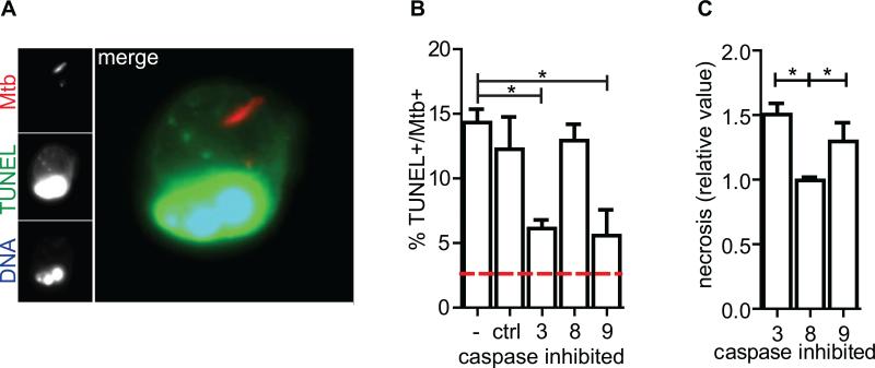
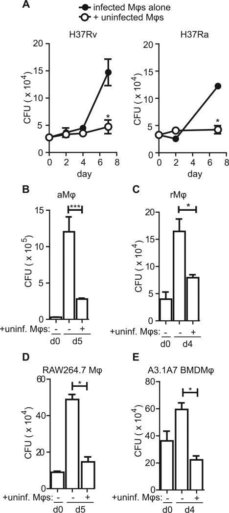
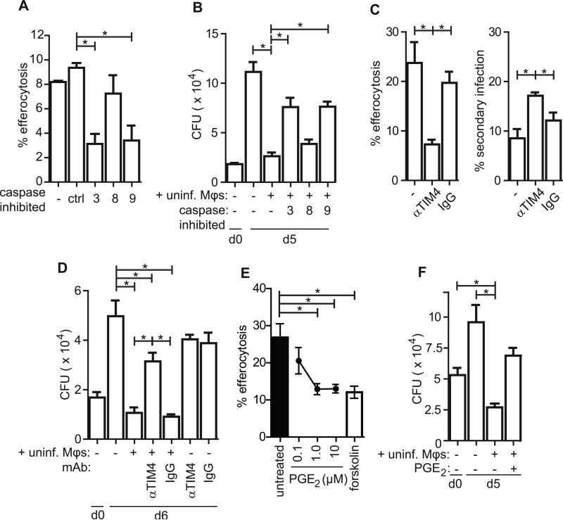
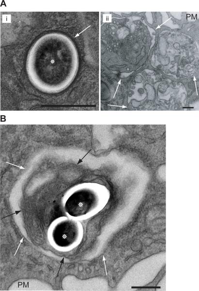
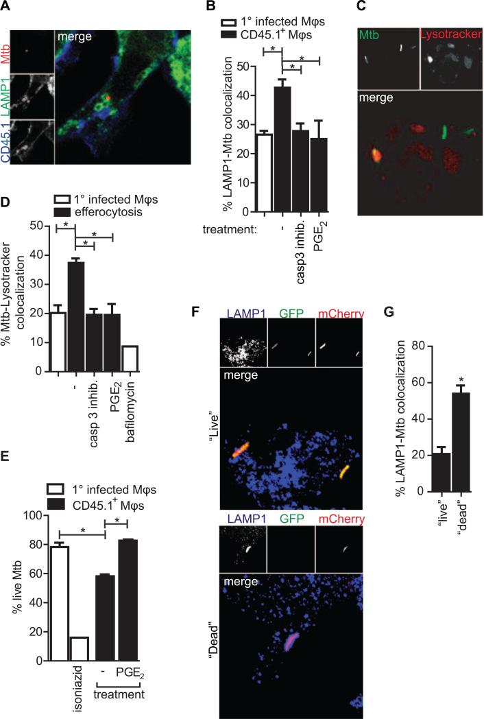
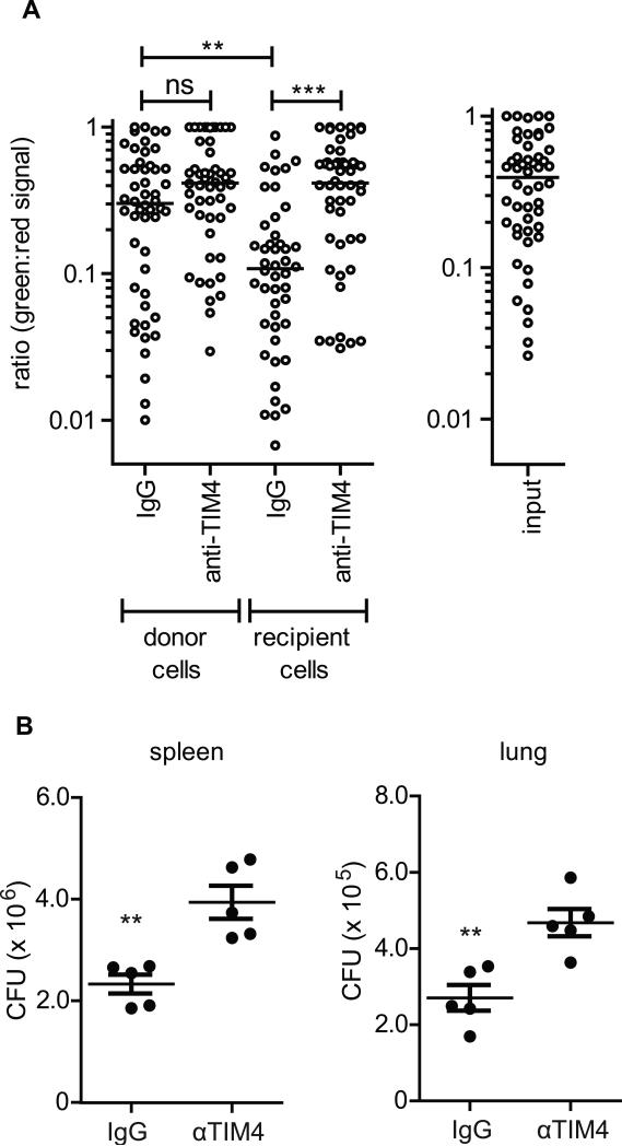
Mycobacterium tuberculosis persists within macrophages in an arrested phagosome and depends upon necrosis to elude immunity and disseminate. Although apoptosis of M. tuberculosis-infected macrophages is associated with reduced bacterial growth, the bacteria are relatively resistant to other forms of death, leaving the mechanism underlying this observation unresolved. We find that after apoptosis, M. tuberculosis-infected macrophages are rapidly taken up by uninfected macrophages through efferocytosis, a dedicated apoptotic cell engulfment process. Efferocytosis of M. tuberculosis sequestered within an apoptotic macrophage further compartmentalizes the bacterium and delivers it along with the apoptotic cell debris to the lysosomal compartment. M. tuberculosis is killed only after efferocytosis, indicating that apoptosis itself is not intrinsically bactericidal but requires subsequent phagocytic uptake and lysosomal fusion of the apoptotic body harboring the bacterium. While efferocytosis is recognized as a constitutive housekeeping function of macrophages, these data indicate that it can also function as an antimicrobial effector mechanism.


Figures

Figure 1
Figure 1. Mtb-infected macrophages undergo apoptosis
(A) Epifluorescence microscopy image of apoptotic Mtb-infected CD11b+ thioglycollate-elicited peritoneal macrophage (pMφ). Mtb (red) TUNEL (green), DNA (UV/pseudocolor blue) and merge. (B) TUNEL+ Mtb-infected pMφs as a percent of total infected pMφs two days post-infection in the presence of 10μM caspase inhibitors or control peptide, ctrl. Red dashed line is the mean percent TUNEL+ of uninfected Mφs. (C) Cell Death ELISA assay for necrosis 2 days post Mtb-infection of pMφs in the presence of 10μM caspase inhibitors relative to infected control-treated Mφs. Data is representative of ≥3 experiments. One way ANOVA with Dunnet's post-test *, p <0.05. Error bars, mean ± SEM.
Figure 2
Figure 2. Efferocytosis of Mtb-infected apoptotic macrophages in vitro and in vivo
(A) Assay for detecting efferocytosis of Mtb-infected apoptotic cells in vitro by confocal microscopy and its three predicted outcomes. (B) Confocal microscopy images of CD45.2+ pMφs dyed with DiO, infected with mCherry-H37Rv and co-cultured with uninfected undyed CD45.1+ pMφs for 16 hours before fixation and visualization. Depicted are the three predicted outcomes of the efferocytosis assay; primary infection (i), secondary infection (ii) and efferocytosis (iii). (C) Efferocytosis seen using the above assay performed with bone marrow-derived macrophages (BMDMφs). Images are representative of >20 experiments. (D) Efferocytosis seen following mCherry-H37Rv infection of DiO-dyed monocyte-derived human macrophages (hMDMφs) and co-culture of uninfected Dextran-649 labeled hMDMφs. (E) Representative images of recovered cells following bronchial alveolar lavage (BAL) 16 hours post transfer of infected pMφs. Images are representative of two experiments. See also Figure S1
Figure 3
Figure 3. Uninfected macrophage co-culture limits bacterial growth
(A,B) pMφs were infected with a 10:1 MOI of H37Rv (A) or H37Ra (B) for four hours before uninternalized bacteria were washed away and uninfected pMφs were added at a ratio of 2:1. Bacterial burden was assessed by CFU enumeration by plating serial dilutions of each condition in quadruplicate at the indicated timepoints. Data is representative of >20 experiments (A, H37Rv) and 3 experiments (B, H37Ra). (C) Alveolar Mφs (aMφs) were infected with an MOI of 10:1 H37Rv for 4 hours before the addition of uninfected aMφs (+unif. Mφs). CFU was enumerated as described on days 0 (d0) and day 5 (d5). Data is representative of 2 experiments. (D) Resident peritoneal Mφs (rMφs) were infected as described with H37Rv before the addition of uninfected rMφs and CFU enumeration on indicated days. Data is representative of 3 experiments. (E) RAW264.7 Mφs were infected with H37Rv as previously described prior to the addition of uninfected Mφs of the same type. CFU was determined. Data is representative of 2 experiments. (F) RAW264.7 Mφs were infected with H37Rv as previously described prior to the addition of uninfected Mφs of the same type. Data is representative of 2 experiments. Error bars ± SEM, p ≤ 0.05, Student's T-test.
Figure 4
Figure 4. Efferocytosis controls Mtb growth
(A) pMφs were infected as described in the confocal microscopy efferocytosis assay in the presence of 10μM caspase inhibitors. Percent efferocytosis is defined as the number of Mφs containing efferocytosed Mtb as a percent of total infected Mφs. Data is representative of 3 experiments. (B) pMφs were infected as described in Fig.4 (see legend), in the presence of 10μM caspase inhibitors. CFU were determined by plating conditions in quadruplicate on days 0 and 5. Data is representative of 3 experiments. (C) rMφs were infected as per the confocal microscopy efferocytosis assay in the presence of 10μg/mL TIM4 blocking antibody 5G3 or control rat IgG1 and efferocytosis and secondary infection was enumerated as above. Data is representative of 3 experiments. (D) rMφs were infected as in Fig.4 in the presence of αTIM4 or control IgG as indicated and CFU were plated in quadruplicate on days 0 and 6 post infection. Data is representative of 4 experiments. (E) Confocal microscopy efferocytosis assay with infected pMφs as previously described. Uninfected pMφs were pre-treated with PGE2 (at indicated concentrations) or forskolin (100μM) for 4 hours prior to washing and addition to the infected pMφ culture. Data is representative of 2 experiments. (F) aMφs were infected according to the efferocytosis assay prior to the addition of uninfected aMφs or PGE2-pretreated aMφs. CFU was determined by plating in quadruplicate on days 0 and 5 post infection. Data is representative of 2 experiments. Error bars ± SEM, p ≤ 0.05, One-way ANOVA with Dunnet's post-test. See also Figure S2.
Figure 5
Figure 5. Mtb are found within vacuous efferocytic phagosomes along with cell debris following uninfected macrophage co-culture
(A) Transmission electron micrograph of (i) Mtb (asterisks) within a pMφ stained with lead citrate. Apoptotic thymocytes co-cultured with pMφs for 1 hour (ii) are engulfed and retained in phagsomes. Note the three thymocyte apoptotic blebs in increasing stages of digestion and disintegration. (B) Mtb within vacuous efferocytic phagosomes inside apoptotic cell blebs after the co-culture of uninfected pMφs with infected pMφs. PM, plasma membrane; scale bar, 1μM; white arrows, phagosome membrane; black arrow, apoptotic bleb membrane. Images are representative from 2 experiments with >1000 Mφs examined on multiple non-serial 60nM sections. See also Figure S3.
Figure 6
Figure 6. Efferocytosis delivers Mtb to the lysosome
(A) Mtb within a LAMP1+ vesicle inside an efferocytic BMDMφ. (B) Quantification of (A), percent of Mtb co-localizing with LAMP1 of total Mtb in primary infected BMDMφs (clear bar) or CD45.1+ BMDMφs (black bars). Uninfected CD45.1+ BMDMφs were untreated, pretreated with PGE2 or added to infected BMDMφs in the presence of caspase 3 inhibitor. Data is representative of 4 experiments. (C) Mtb within acidic vesicles pMφs. (D) Quantification of (C) showing percent Mtb-Lysotracker Red colocalization of total GFP-Mtb in primary infected pMφs or following efferocytosis and the depicted treatments. Bafilomycin was added at 1μM for 2 hours before and during Lysotracker staining. Data is representative of 2 experiments. (E) Percent “Live” Mtb of total Mtb counted in primary infected pMφs (clear bar) or CD45.1+ pMφs following co-culture with infected macrophages. Isoniazid was added for 24 hours. Data is representative of two experiments. Error bars ± SEM, p ≤ 0.05, One-way ANOVA with Dunnet's post-test. (F) “Live” (top) Mtb do not co-localize with LAMP1 as frequently as “Dead” (bottom) Mtb in bone marrow-derived macrophages (BMDMφs). (G) Quantification of (F). Percent LAMP1 colocalization with “Live” or “Dead” Mtb of total “Live” or “Dead” Mtb in BMDMφs. Data is representative of two experiments. Error bars ± SEM, p ≤ 0.05. Student's T-test. See also Figure S4.
Figure 7
Figure 7. Efferocytosis controls Mtb in vivo
(A) Mtb is less viable following efferocytosis in vivo. Live/Dead-H37Rv 5LO-/- infected pMφs were intraperitoneally transferred into CD45.1 mice with IgG or aTIM4 antibody. Sixteen hours later cells were removed and treated with 200ng/mL anhydrous tetracycline to induce GFP expression. Mtb viability was ascertained by measuring the ratio of GFP:mCherry expression for each bacterium. Input is expression in untransferred infected pMφs. Data is representative of 2 experiments. Median, p ≤ 0.001. Mann-Whitney test. (B) Bacterial burden in spleen and lung of Rag-/- mice two weeks post intravenous transfer of H37Rv-infected 5LO-/- pMφs and intraperitoneal administration of IgG or aTIM4. Data is representative of one experiment. Error bars ± SEM, p ≤ 0.001. Student's T-test. See also Figure S5.

Comment in

Similar articles

Cited by

References

    1. Albacker LA, Karisola P, Chang YJ, Umetsu SE, Zhou M, Akbari O, Kobayashi N, Baumgarth N, Freeman GJ, Umetsu DT, et al. TIM-4, a receptor for phosphatidylserine, controls adaptive immunity by regulating the removal of antigen-specific T cells. J Immunol. 2010;185:6839–6849. - PMC - PubMed
    1. Armstrong JA, Hart PD. Response of cultured macrophages to Mycobacterium tuberculosis, with observations on fusion of lysosomes with phagosomes. The Journal of experimental medicine. 1971;134:713–740. - PMC - PubMed
    1. Aronoff DM, Canetti C, Peters-Golden M. Prostaglandin E2 inhibits alveolar macrophage phagocytosis through an E-prostanoid 2 receptor-mediated increase in intracellular cyclic AMP. J Immunol. 2004;173:559–565. - PubMed
    1. Carpenter AE, Jones TR, Lamprecht MR, Clarke C, Kang IH, Friman O, Guertin DA, Chang JH, Lindquist RA, Moffat J, et al. CellProfiler: image analysis software for identifying and quantifying cell phenotypes. Genome Biol. 2006;7:R100. - PMC - PubMed
    1. Chen M, Divangahi M, Gan H, Shin DS, Hong S, Lee DM, Serhan CN, Behar SM, Remold HG. Lipid mediators in innate immunity against tuberculosis: opposing roles of PGE2 and LXA4 in the induction of macrophage death. The Journal of experimental medicine. 2008;205:2791–2801. - PMC - PubMed

Publication types