. 2012 Sep 13;12(3):334-45.
doi: 10.1016/j.chom.2012.07.013.

A neuron-specific role for autophagy in antiviral defense against herpes simplex virus

Affiliations

A neuron-specific role for autophagy in antiviral defense against herpes simplex virus

Brian Yordy et al. Cell Host Microbe. .

Abstract

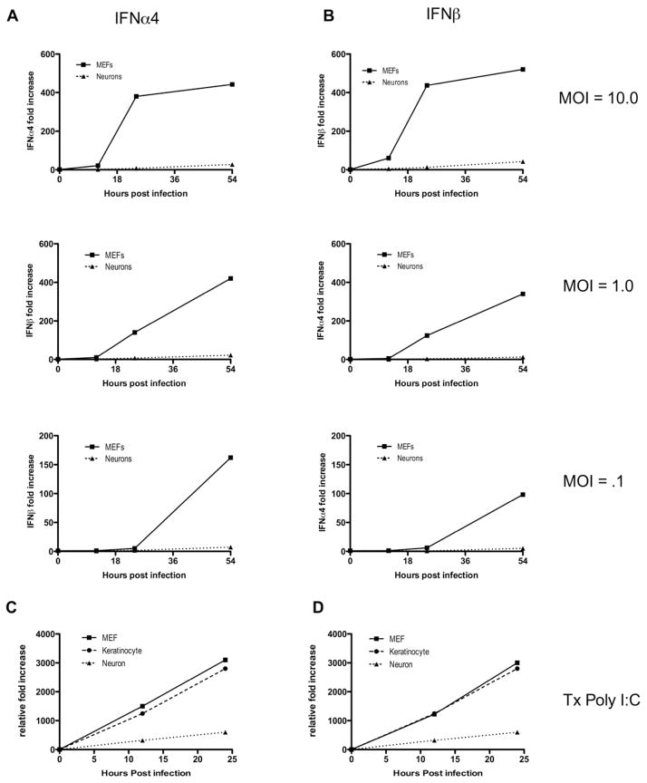
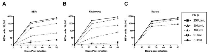
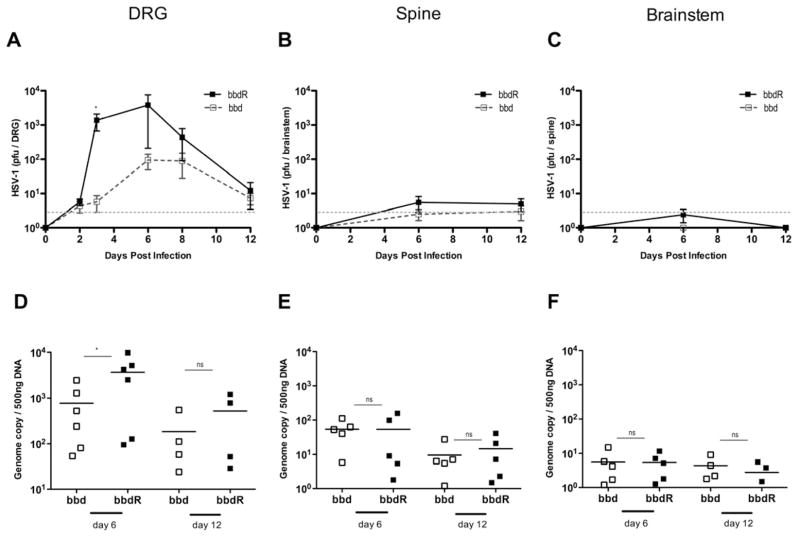
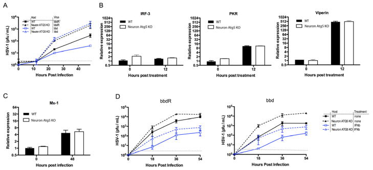
Type I interferons (IFNs) are considered to be the universal mechanism by which viral infections are controlled. However, many IFN-stimulated genes (ISGs) rely on antiviral pathways that are toxic to host cells, which may be detrimental in nonrenewable cell types, such as neurons. We show that dorsal root ganglionic (DRG) neurons produced little type I IFNs in response to infection with a neurotropic virus, herpes simplex type 1 (HSV-1). Further, type I IFN treatment failed to completely block HSV-1 replication or to induce IFN-primed cell death in neurons. We found that DRG neurons required autophagy to limit HSV-1 replication both in vivo and in vitro. In contrast, mucosal epithelial cells and other mitotic cells responded robustly to type I IFNs and did not require autophagy to control viral replication. These findings reveal a fundamental difference in the innate antiviral strategies employed by neurons and mitotic cells to control HSV-1 infection.


Conflict of interest statement

Authors declare no conflicts of interest.

Figures

Figure 1
Figure 1. Neurons are impaired in their capacity to respond to viral stimulation and induce type I IFNs
(A, B) Mouse embryonic fibroblasts (MEFs) and neurons were infected with HSV1-GFP at MOIs of 0.1, 1.0 and 10.0. RNA was isolated from all cells at 0, 12, 24, and 54 hours post-infection. Relative fold increases in IFN-α4 (A) and IFN-β (B) production were determined by quantitative RT-PCR of isolated RNA relative to the housekeeping gene hprt. (C, D) MEFs, keratinocytes, and neurons were transfected with Poly I:C (1.0 μg/mL). RNA was isolated from all cells at the time points indicated, and fold increases of IFN-α4 (C) and IFN-β (D) relative to hprt were determined by RT-qPCR. Data are representative of three independent experiments. See also Figure S1.
Figure 2
Figure 2. Neurons have impaired type I IFN-dependent anti-HSV-1 protection
(A–C) Primary keratinocytes, MEFs, and neurons were pre-treated for 18 hours with the indicated amount of IFN-β. Following pre-treatment, all cells were infected with 0.5 MOI of HSV1-GFP. At the time points indicated, cells were isolated and analyzed for GFP expression by flow cytometry. Data are shown as number of infected cells (GFP+) per 10,000 cell events for MEFs (A), keratinocytes (B), and neurons (C). Data are representative of four independent experiments. See also Figure S2.
Figure 3
Figure 3. Type I interferon primes mitotic cells, but not neurons, for cell death
(A) MEFs, and neurons were pre-treated for 18 hours with the indicated amount of IFN-β. All cells were then transfected with Poly I:C (1.0 μg/mL) for one hour. At the time points indicated, MEFs and neurons were isolated, stained with trypan blue and viable cells were counted by hematocytometer (top panel). MEFs and neurons were isolated, stained with Annexin V and 7-AAD, and analyzed by flow cytometry (bottom panel). Data are representative of three independent experiments. (B) MEFs and neurons were treated with staurosporine and isolated, stained with trypan blue, and counted by hematocytometer at the time points indicated. Error bars represent SEM. See also Figure S3.
Figure 4
Figure 4. Viral inhibition of autophagy in an infected, non-epithelial cell type is required for HSV-1-induced disease
(A–D) Keratinocytes and total liver were isolated from ATG5flox.flox Krt14cre+ (KO) and ATG5flox.flox Krt14cre- (WT) mice. ATG5 mRNA expression relative to levels in KO is shown for keratinocytes (A) and liver (C). Western blot analysis of LC3-I and LC3-II from isolated keratinocytes (B) and total liver (D) is shown. Loading control (β-actin) for each sample is shown below. n > 2 for all genotypes and data are representative of two independent experiments. (E–H) Depo-provera treated Atg5 flox/flox x Krt14-cre Tg (KC-Atg5 KO) and Atg5 flox/flox Krt14cre(−) (WT) littermates were intravaginally infected with 5,000 pfu of either HSV-1 bbd or the rescue virus bbdR. (E) Vaginal washes were collected daily and viral titers were measured. Mice were weighed (F), clinically scored (G), and monitored for survival (H) every 24 hours post infection. Error bars represent SEM of n > 12 mice per group. **p < .001; ***; p < .0005 mortality of bbdR infected mice relative to bbd infection. Data are pooled from three independent experiments. See also Figure S4.
Figure 5
Figure 5. Neuro-attenuation of bbd is independent of T-cell responses
(A–C) Depo-provera treated WT C57BL/6 mice were intravaginally infected with 20,000 pfu of HSV-1 bbd or the rescue mutant bbdR, and were analyzed on days six and eight post infection. Total number of lymphocytes (A), and CD4+ T-cells (B) from the iliac and lingual draining lymph nodes is depicted. (C) Isolated CD4+ T-cells were co-cultured with naïve splenocytes in the presence of heat-inactivated HSV-1. IFN-γ secretion from CD4+ T cells was measured by ELISA. (D–G) CD4−/−, CD8−/−, and WT control C57BL/6 mice were intravaginally infected with 2,000 pfu of the indicated strain of HSV-1. Vaginal washes of CD4−/− (D) and CD8−/− (F) mice were collected daily and viral titers were measured in Vero cells. CD4−/− (E) and CD8−/− (G) mice were monitored for survival every 24 hours post infection. Error bars represent SEM of n > 5 mice per group. Data are pooled from four (A–C) or three (D–F) independent experiments. See also Figure S5.
Figure 6
Figure 6. Autophagy is required to restrict neuronal HSV-1 replication
Depo-provera treated wild-type C57BL/6 mice were infected with 2 × 10⁴ pfu of the indicated strain of HSV-1. Dorsal root ganglia, spinal cord, and brainstem were isolated at the time points indicated, homogenized. Viral titers were measured in Vero cells (A–C) and genome copy of isolated DNA was determined by qPCR (D–F). Error bars represent SEM (A–C) of n > 5, n>4 (D–F). Gray dotted line indicates limit of detection. Data are pooled from three independent experiments. See also Figure S6.
Figure 7
Figure 7. Autophagy and type I IFNs restrict HSV-1 replication in neurons in parallel
Primary neurons were isolated from Atg5 flox/flox x Nestin-cre Tg (Neuron Atg5 KO) and Atg5 flox/flox Nestin-cre(−) (WT) littermates. Neurons were infected at the indicated MOI with either bbd or bbdR HSV-1, at MOI of 0.01 (A), or at MOI of 0.1 with or without treatment with 250 U/ml of IFN-β 18 hours prior to infection (D). (A, D) Supernatants were collected at points indicated and viral titers were measured. (B & C) RNA isolated from neurons treated with 1,000 U/ml of IFN-β alone (B), or followed by HSV-1 infection for additional 48 hours was used to measure expression of indicated ISG by RT-qPCR. Error bars represent SEM and data are representative of two to four independent experiments. Gray dotted line indicates limit of detection. See also Figure S7.

Similar articles

Cited by

References

    1. Alexander DE, Leib DA. Xenophagy in herpes simplex virus replication and pathogenesis. Autophagy. 2008;4:101–103. - PMC - PubMed
    1. Alexander DE, Ward SL, Mizushima N, Levine B, Leib DA. Analysis of the role of autophagy in replication of herpes simplex virus in cell culture. Journal of virology. 2007;81:12128–12134. - PMC - PubMed
    1. Alirezaei M, Kiosses WB, Flynn CT, Brady NR, Fox HS. Disruption of neuronal autophagy by infected microglia results in neurodegeneration. PLoS One. 2008;3:e2906. - PMC - PubMed
    1. Barber GN. Host defense, viruses and apoptosis. Cell Death and Differentiation. 2001;8:113. - PubMed
    1. Brot MD, Rall GF, Oldstone MBA, Koob GF, Gold LH. Deficits in discriminated learning remain despite clearance of long-term persistent viral infection in mice. Journal of NeuroVirology. 1997;3:265. - PubMed

Publication types

Substances

LinkOut - more resources