

Global prefrontal and fronto-amygdala dysconnectivity in bipolar I disorder with psychosis history
- PMID: 22980587
- PMCID: PMC3549314
- DOI: 10.1016/j.biopsych.2012.07.031
Global prefrontal and fronto-amygdala dysconnectivity in bipolar I disorder with psychosis history
Abstract
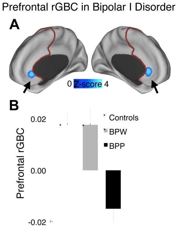
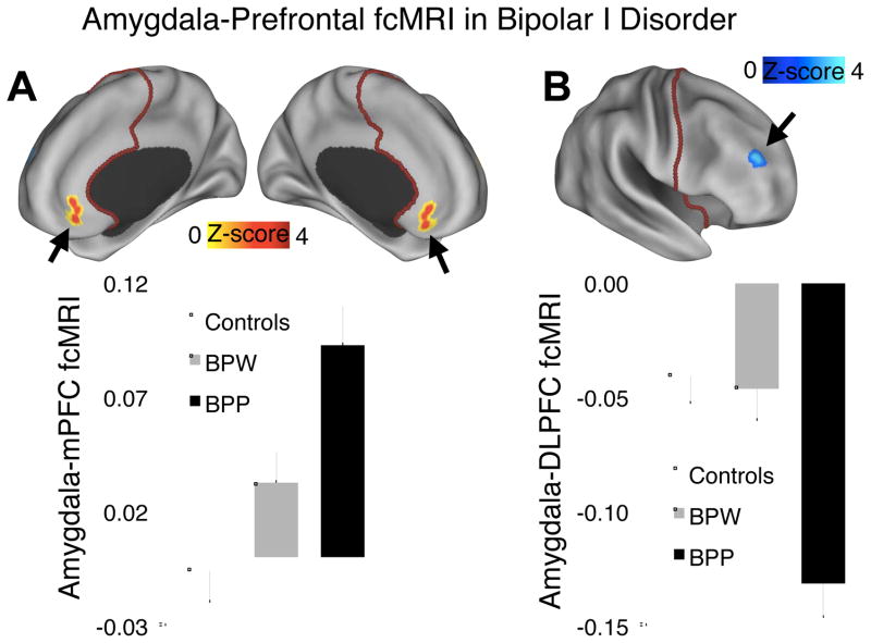
Background: Pathophysiological models of bipolar disorder postulate that mood dysregulation arises from fronto-limbic dysfunction, marked by reduced prefrontal cortex (PFC) inhibitory control. This might occur due to both disruptions within PFC networks and abnormal inhibition over subcortical structures involved in emotional processing. However, no study has examined global PFC dysconnectivity in bipolar disorder and tested whether regions with within-PFC dysconnectivity also exhibit fronto-limbic connectivity deficits. Furthermore, no study has investigated whether such connectivity disruptions differ for bipolar patients with psychosis history, who might exhibit a more severe clinical course.
Methods: We collected resting-state functional magnetic resonance imaging at 3T in 68 remitted bipolar I patients (34 with psychosis history) and 51 demographically matched healthy participants. We employed a recently developed global brain connectivity method, restricted to PFC (rGBC). We also independently tested connectivity between anatomically defined amygdala and PFC.
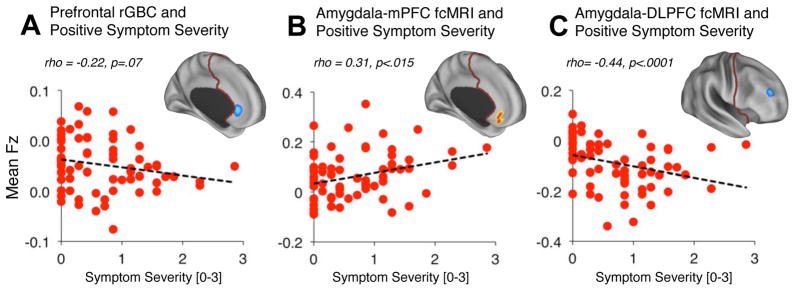
Results: Bipolar patients exhibited reduced medial prefrontal cortex (mPFC) rGBC, increased amygdala-mPFC connectivity, and reduced connectivity between amygdala and dorsolateral PFC. All effects were driven by psychosis history. Moreover, the magnitude of observed effects was significantly associated with lifetime psychotic symptom severity.
Conclusions: This convergence between rGBC, seed-based amygdala findings, and symptom severity analyses highlights that mPFC, a core emotion regulation region, exhibits both within-PFC dysconnectivity and connectivity abnormalities with limbic structures in bipolar illness. Furthermore, lateral PFC dysconnectivity in patients with psychosis history converges with published work in schizophrenia, indicating possible shared risk factors. Observed dysconnectivity in remitted patients suggests a bipolar trait characteristic and might constitute a risk factor for phasic features of the disorder.
Copyright © 2013 Society of Biological Psychiatry. Published by Elsevier Inc. All rights reserved.
Figures
Comment in
-
Imaging symptoms and syndromes: similarities and differences between schizophrenia and bipolar disorder.Biol Psychiatry. 2013 Mar 15;73(6):495-6. doi: 10.1016/j.biopsych.2013.01.015. Biol Psychiatry. 2013. PMID: 23438631 No abstract available.
References
-
- Chen C-H, Suckling J, Lennox BR, Ooi C, Bullmore ET. A quantitative meta-analysis of fMRI studies in bipolar disorder. Bipolar Disord. 2011;13:1–15. - PubMed
-
- Altshuler L, Bookheimer S, Proenza MA, Townsend J, Sabb F, Firestine A, et al. Increased amygdala activation during mania: a functional magnetic resonance imaging study. Am J Psychiatry. 2005;162:1211–1213. - PubMed
-
- Blumberg HP, Kaufman J, Martin A, Whiteman R, Zhang JH, Gore JC, et al. Amygdala and hippocampal volumes in adolescents and adults with bipolar disorder. Arch Gen Psychiatry. 2003;60:1201–1208. - PubMed
Publication types
MeSH terms
Grants and funding
LinkOut - more resources
Full Text Sources
Other Literature Sources
Medical
Miscellaneous
