. 2012 Sep 14;150(6):1264-73.
doi: 10.1016/j.cell.2012.08.020.

Long-distance growth and connectivity of neural stem cells after severe spinal cord injury

Affiliations

Long-distance growth and connectivity of neural stem cells after severe spinal cord injury

Paul Lu et al. Cell. .

Abstract

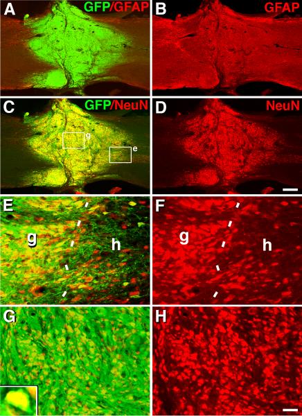
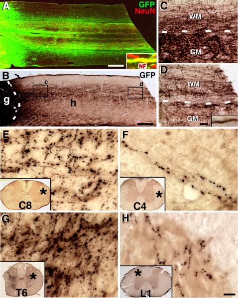
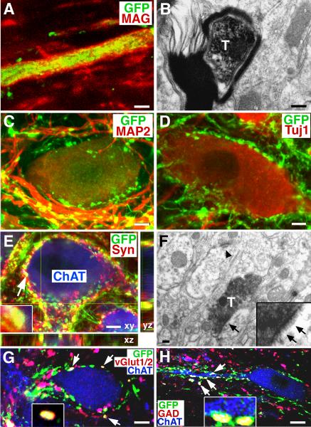
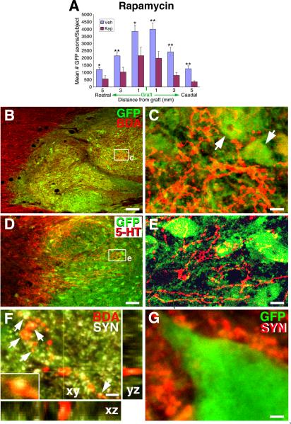
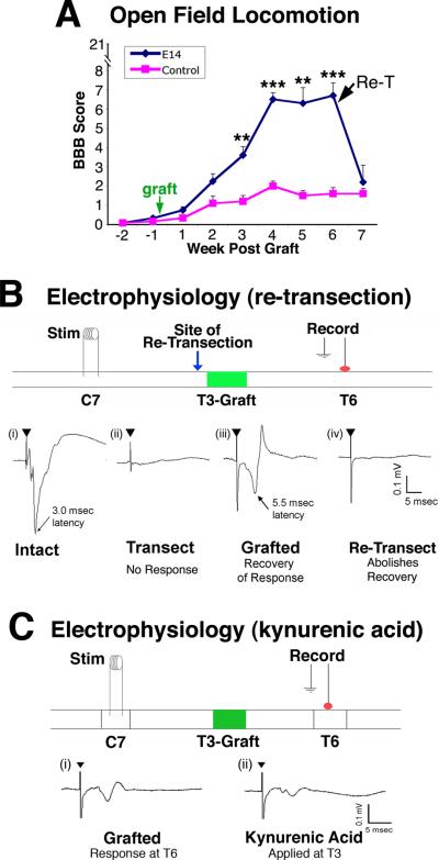
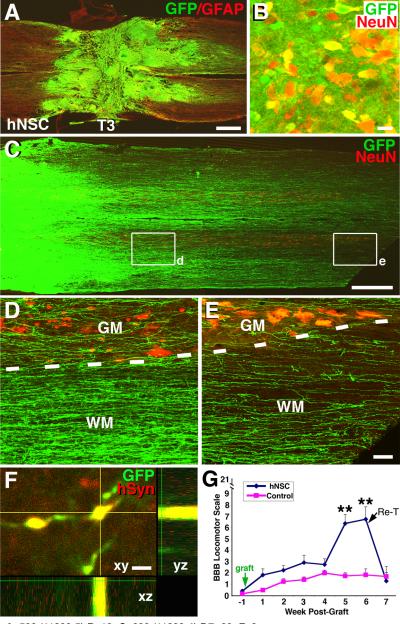
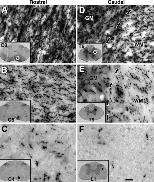
Neural stem cells (NSCs) expressing GFP were embedded into fibrin matrices containing growth factor cocktails and grafted to sites of severe spinal cord injury. Grafted cells differentiated into multiple cellular phenotypes, including neurons, which extended large numbers of axons over remarkable distances. Extending axons formed abundant synapses with host cells. Axonal growth was partially dependent on mammalian target of rapamycin (mTOR), but not Nogo signaling. Grafted neurons supported formation of electrophysiological relays across sites of complete spinal transection, resulting in functional recovery. Two human stem cell lines (566RSC and HUES7) embedded in growth-factor-containing fibrin exhibited similar growth, and 566RSC cells supported functional recovery. Thus, properties intrinsic to early-stage neurons can overcome the inhibitory milieu of the injured adult spinal cord to mount remarkable axonal growth, resulting in formation of new relay circuits that significantly improve function. These therapeutic properties extend across stem cell sources and species.


Figures

Figure 1
Figure 1. Survival, Filling and Differentiation of Neural Stem Cell Grafts in T3 Complete Transection Site
(A–B) Overview of GFP and GFAP fluorescent immunolabeling in a horizontal section demonstrates excellent graft survival, integration and filling of T3 complete transection site, seven weeks post-grafting. (C–D) GFP and NeuN labeling confirm extensive neuronal differentiation/maturation of grafted rat neural stem cells. (E–F) Higher magnification from c showing excellent integration and transition from host (h) neurons to grafted (g) neurons (dashed lines) (E: GFP, NeuN; F, NeuN alone). (G–H) Higher magnification from center of graft showing high density of NeuN-labeled neurons (inset) (G: GFP, NeuN; H, NeuN alone). Scale bar: A–D, 320 μm; E–H, 48 μm. Also see Figure S1 and S2.
Figure 2
Figure 2. Extensive Long-Distance Axonal Outgrowth from Neural Stem Cell Grafts
(A) GFP and NeuN immunolabeling reveals that GFP-expressing neural stem cell grafts robustly extend axons into the host spinal cord rostral and caudal to the T3 complete transection site (caudal shown) over the 12mm length of the horizontal section. Extensive regions of the host spinal cord contain graft-derived projections in white matter and gray matter. Inset shows that GFP-labeled projections arising from grafts express neurofilament (NF), confirming their identity as axons. (B) Light-level GFP immunolabeling also clearly reveals the density and distribution of axons extending from the lesion site. Boxes are shown at higher magnification in C, D. (C) Dense numbers of GFP-labeled axons are present in host white matter (WM) and gray matter (GM) 2mm caudal to the lesion, and (D) large numbers of axons remain at the end of the block of tissue, 6mm caudal to the lesion. (E) A high density of GFP-labeled axons is present in the lateral portion of C8, three spinal segments above the lesion, and (F) remain detectable at C4, 7 spinal segments above the lesion. Asterisk indicates locations of higher magnification view. (G) GFP-labeled axons also extend in dense numbers caudally to T6 white matter and gray matter (shown) and (H) caudally to L1. Overall, axons extend at least 25 mm in each direction. Scale bar: A–B, 600 μm; B–C, 40 μm; D–G, 10 μm. Also see Figure S3 and S4.
Figure 3
Figure 3. Myelination, Synapse Formation and Expression of Neurotransmitters
(A–B) Graft-derived, GFP-labeled axons are myelinated in many cases, (red, myelin-associated glycoprotein, MAG, confirmed by electron microscopy (T, transplanted, GFP-labeled axon). (C) GFP-expressing axon terminals are closely associated with host MAP-2-expressing neurons and dendrites, and (D) host Tuj1-expressing neuronal somata. (E) A z-stack image triple labeled for GFP, synaptophysin (Syn, inset), and ChAT, indicating co-association of graft-derived axons with a synaptic marker in direct association with host motor neurons (arrowhead indicates one of several examples). (F) Electron microscopy confirms that DAB-labeled GFP-expressing axon terminals form synapses (arrows) with host dendrites. Arrowhead indicates a separate, host-host synapse. (G–H) Expression of vGlut1/2 or GAD65 by GFP-labeled axons (arrows and insets) in close association with host motor (ChAT-labeled) neurons. Scale bar: A, 3μm; B, 200nm; C–E, 8μm; F, 200nm; G, 7μm; H, 6μm.
Figure 4
Figure 4. Rapamycin Reduces Axon Growth; Host Axons Innervate Grafts
(A) Treatment with the mTOR inhibitor rapamycin significantly reduced outgrowth of graft-derived axons (*p<0.05, **p<0.01; see also Suppl. Fig 5). Veh, vehicle; Rap, rapamycin treated. Data are represented as mean ± SEM. (B–C) Host reticulospinal axons labeled with BDA regenerate into GFP-expressing neural stem cell grafts in site of T3 complete transection (C, from boxed area of panel B; arrows indicate GFP-labeled grafted cells with neuronal morphology). (D–E) Host serotonergic axons immunolabeled for 5-HT regenerate into GFP expressing E14 neural stem cell grafts in lesion site. (F) Bouton-like structures on host reticulospinal axons regenerating into graft co-localize with synaptophysin in a Z-stack image (arrows). (G) Host synaptophysin terminals (red) are closely apposed to GFP-labeled grafted cells exhibiting neuronal morphology. Scale bar: B, 64μm; C, 12μm; D, 70 μm; E, 10 μm; F–G, 2μm. .
Figure 5
Figure 5. Functional and Electrophysiological Improvement after T3 Complete Transection
(A) Hindlimb locomotion: BBB scores after T3 complete transection show significant improvement in subjects that received neural stem cell grafts (NSC, n=6) compared to lesioned controls (n=6). Re-transection (arrow, Re-T) at rostral interface of graft with host abolishes functional improvements when assessed one week later (**p<0.01, ***p<0.001). Data are represented as mean ± SEM. (B) Electrophysiological transmission across the T3 complete lesion site: (i) In intact animals, stimulation at C7 evoked a short latency (~3.0 msec), large amplitude response at T6. (ii) Transection of the cord at T3 completely abolished this response. (iii) In 4 of 6 lesion/grafted animals, recovery of an evoked response of prolonged latency (~5.5 msec) was observed. (iv) Re-transection of the spinal cord at T3, just rostral to the graft (green arrow), abolished the recovered evoked response. (C) Kynurenic acid (50 mM), a blocker of excitatory synapses, was applied to the graft to determine whether excitatory transmission across synapses in the graft was required to detect responses at T6. Indeed, kynurenic acid application substantially reduced the amplitude but not the latency of the evoked response. Also see Figure S6.
Figure 6
Figure 6. Human Neural Stem Cell Grafts in Fibrin Matrices after T3 Transection: Long-Distance Axonal Growth, Connectivity and Functional Improvement
(A) GFP-expressing 566RSC human neural stem cells (NSCs) survive and completely fill T3 complete transection site, 7 weeks post-grafting (GFP, green; GFAP, red). (B) GFP and NeuN labeling reveals that many grafted human neural stem cells express mature neuronal markers. Field is from graft center. (C–E)GFP-expressing human neurons robustly extend axons into the host spinal cord caudal and rostral to the complete T3 transection site (caudal shown). Axons extend through both white matter (WM) and gray matter (GM). (F) GFP-labeled human axon terminals express human-specific synaptophysin (hSyn) in host gray matter. Site shown is 7 mm caudal to lesion. Scale bar: A, 500 μm; B, 10 μm; C, 600 μm; D–E, 60 μm; F 3 μm. (G) There is significant functional improvement after human NSC grafts to sites of T3 complete transection (**p<0.01). Re-transection (arrow, Re-T) at rostral interface of graft completely abolishes functional recovery assessed one week later, indicating that host inputs are required to improve function. Data are represented as mean ± SEM.
Figure 7
Figure 7. Dense Axonal Outgrowth from Implants of Human 566RSC Neural Stem Cells
(A) Very large numbers of GFP-labeled axons extend into host spinal cord rostral to T3 transection site, 7 weeks post-grafting. C8 segment shown. (B–C) GFP-labeled axons are present in C6 and C4 white matter, five and seven spinal segments above the lesion, respectively. Asterisks indicate location of higher magnification views. (D–F) GFP-labeled axons also extend in high density caudal to T6 and T9, and are present in both white matter (WM) and gray matter (GM, dashed lines indicate interface). (F) Axons continue to extend caudally to L1. Overall, axons extend at least 25 mm in each direction in all subjects. Scale bar: 15 μm.

Comment in

Similar articles

Cited by

References

    1. Bamber NI, Li H, Aebischer P, Xu XM. Fetal spinal cord tissue in miniguidance channels promotes longitudinal axonal growth after grafting into hemisected adult rat spinal cords. Neural Plast. 1999;6:103–121. - PMC - PubMed
    1. Baska KM, Manandhar G, Feng D, Agca Y, Tengowski MW, Sutovsky M, Yi YJ, Sutovsky P. Mechanism of extracellular ubiquitination in the mammalian epididymis. J Cell Physiol. 2008;215:684–696. - PubMed
    1. Basso DM, Beattie MS, Bresnahan JC. Graded histological and locomotor outcomes after spinal cord contusion using the NYU weight-drop device versus transection. Exp Neurol. 1996;139:244–256. - PubMed
    1. Bonner JF, Connors TM, Silverman WF, Kowalski DP, Lemay MA, Fischer I. Grafted neural progenitors integrate and restore synaptic connectivity across the injured spinal cord. J Neurosci. 2011;31:4675–4686. - PMC - PubMed
    1. Boulis NM, Federici T, Glass JD, Lunn JS, Sakowski SA, Feldman EL. Translational stem cell therapy for amyotrophic lateral sclerosis. Nat Rev Neurol. 2011;8:172–176. - PubMed

Publication types

Substances