. 2012 Sep 27;2(3):553-67.
doi: 10.1016/j.celrep.2012.08.002. Epub 2012 Sep 13.

Identification of the hemogenic endothelial progenitor and its direct precursor in human pluripotent stem cell differentiation cultures

Affiliations

Identification of the hemogenic endothelial progenitor and its direct precursor in human pluripotent stem cell differentiation cultures

Kyung-Dal Choi et al. Cell Rep. .

Abstract

Hemogenic endothelium (HE) has been recognized as a source of hematopoietic stem cells (HSCs) in the embryo. Access to human HE progenitors (HEPs) is essential for enabling the investigation of the molecular determinants of HSC specification. Here, we show that HEPs capable of generating definitive hematopoietic cells can be obtained from human pluripotent stem cells (hPSCs) and identified precisely by a VE-cadherin(+)CD73(-)CD235a/CD43(-) phenotype. This phenotype discriminates true HEPs from VE-cadherin(+)CD73(+) non-HEPs and VE-cadherin(+)CD235a(+)CD41a(-) early hematopoietic cells with endothelial and FGF2-dependent hematopoietic colony-forming potential. We found that HEPs arise at the post-primitive-streak stage of differentiation directly from VE-cadherin-negative KDR(bright)APLNR(+)PDGFRα(low/-) hematovascular mesodermal precursors (HVMPs). In contrast, hemangioblasts, which are capable of forming endothelium and primitive blood cells, originate from more immature APLNR(+)PDGFRα(+) mesoderm. The demarcation of HEPs and HVMPs provides a platform for modeling blood development from endothelium with a goal of facilitating the generation of HSCs from hPSCs.


Figures

Figure 1
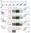
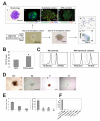
Figure 1. Characterization of major subsets of VE-cadherin+ cells generated from hESCs after 5 days of coculture on OP9
(A) Kinetics of VE-cadherin and CD31 expression in differentiated H1 hESCs. (B) Characterization of endothelial and hematopoietic CFC potentials of freshly isolated day 5 VE-cadherin+ subsets. Histograms represent the expression of typical endothelial molecules by indicated cell subsets. AcLDL histograms show flow cytometric profiles of cells incubated with AcLDL at 370C (AcLDL uptake; black histogram) versus 40C (AcLDL binding control; gray histogram). (i) Wright-stained cytospins demonstrate morphology of isolated cells (bar = 20 μm). Endothelial culture panels show (ii) phase contrast images (bar = 400 μm), (iii) immunofluorescent analysis (bar = 100 μm), and (iv) tube formation (bar = 400 μm). Hematopoietic CFC potential of sorted day 5 VE-cadherin+ subsets evaluated in serum-free Methocult (SF) supplemented with FGF2, SCF, IL6, IL3 and EPO and in standard serum-containing GF+ H4435 MethoCult. Error bars are means ± SE of three experiments. See also Figure S1 and S3.
Figure 2
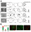
Figure 2. Stroma-dependent hematopoietic and endothelial potential of day 5 VE-cadherin+ subsets
(A) Phase contrast images of cultures (bar = 400 μm), flow cytometric analysis and hematopoietic CFC potential (GF+ H4435 serum-containing MethoCult) are shown. Error bars are means ± SE of three experiments. The numbers show mean counts for CFC-GEMM. (B) A single-cell deposition assay to detect the frequency of endothelial, hematopoietic, and bipotential hematoendothelial progenitors. Immunofluorescent staining of clusters formed by single cells after 10-12 days of culture on OP9 using CD43 and VE-cadherin antibodies is shown (bar = 100 μm). Graph shows the frequency of each type of progenitor as a percentage of cluster-containing wells versus total cell-deposited wells. Error bars are means ± SE of three experiments.
Figure 3
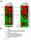
Figure 3. Gene expression profiling of undifferentiated H1 hESCs, differentiated H1 hESC cell populations, and human umbilical endothelial cells
(A) Heat maps of selected genes associated with endothelial and hematopoietic cells, epithelial-mesenchymal transition (EMT), lateral plate/extraembryonic (EE) mesoderm, and primitive streak. HB cores are endothelial intermediates formed by hemangioblasts in serum-free clonogenic medium. EC p2 are second passage of endothelial cells obtained from day 8 CD31+CD43 differentiated H1 cells. HUVEC are human umbilical vein endothelial cells. See Table 1 for other abbreviations. The gene expression levels are estimated in terms of “transcripts per million”. (B) Pearson correlation analysis of global gene expression. See also Figure S4.
Figure 4
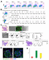
Figure 4. Phenotypic and functional characterization of major subsets of day 4 hESC-derived mesodermal cells
(A) Kinetics of expression of APLNR and KDR in differentiated H1 hESCs. (B) Flow cytometric analysis of day 4 hESC-derived mesodermal cells that were first magnetically sorted for the KDR+ population. (C) Endothelial and hematopoietic differentiation potential of indicated day 4 mesodermal subsets. (i) Wright-stained cytospins of freshly isolated cells (bar = 20 μm). Endothelial culture panels show (ii) phase contrast images (bar = 400 μm), (iii) immunofluorescent analysis (bar = 100 μm), and (iv) tube formation (bar = 400 μm) by KbrA+P cells. (v) shows phase contrast image of KbrA+P cell cultures on OP9. Flow cytometric analysis shows a developmental potential of KDR subsets after secondary OP9 coculture. Hematopoietic CFC potential of KDR subsets was evaluated after secondary coculture on OP9 using standard H4435 GF+ serum-containing MethoCult. Error bars are means ± SE of three experiments. (D) Confocal images of hemtoendothelial clusters to demonstrate the early stages of endothelial-hematopoietic transition; bar = 50 μm. (E) A single-cell deposition assay to detect the frequency of endothelial (E), hematopoietic (H), and bipotential hematoendothelial (HE) progenitors within KbrA+P HVMPs. Graph shows the frequency of each type of progenitor as a percentage of cluster-containing wells versus total cell-deposited wells. Error bars are means ± SE of three experiments. See also Figure S5.
Figure 5
Figure 5. Characterization of BL-CFCs
(A) Hematopoietic and endothelial potential of HB colonies selected at day 3 (core stage; bar = 50 μm) and day 12 (mature blast colony; bar = 100 μm) of clonogenic culture. Top panels show (i) Wright-stained cytospins (bar = 100 μm), (ii) VE-cadherin and AcLDL staining (bar = 50 μm), (iii) endothelial culture (bar = 100 μm), and (iv) OP9 coculture (bar = 100 μm) of HB cores. Left panels show (v) cytospins (bar = 20 μm) and (vi) flow cytometric analysis of mature HB colony. (B) The effect of adding apelin-12 on BL colony formation. Error bars are means ± SE of six experiments. (C) Flow cytometric analysis of expression of adult and embryonic hemoglobins in HB colonies and erythroid colonies derived from V+73235 HEPs after coculture on OP9. (D) Morphology of typical hematopoietic colonies generated from day 12 BL colonies after replating them into H4436 serum-free methylcellulose clonogenic medium containing hematopoietic cytokines; bars = 200 μm. (E) Frequency of hematopoietic colonies formed after replating of HB colonies into serum-free hematopoietic CFC medium. Left panel shows results of replating 20 individual colonies; error bars are means ± SE of three experiments. Right panel shows results of replating 200 blast colonies. (F) BL-CFC potential of indicated cell subsets detected using FGF2 and apelin 12-containing clonogenic medium. Error bars represent standard error of three experiments. See also Figure S6.
Figure 6
Figure 6. Distinct stages of angiohematopoietic differentiation from hPSCs in coculture with OP9
hESC coculture with OP9 induces mesendodermal differentiation. The first cells with angiohematopoietic potential arise within EMHlin APLNR+PDGFRα+ mesoderm. These cells have the potential to form BL (HB) colonies, which can be specifically detected in serum-free semisolid medium containing FGF2 and apelin. Development of BL colonies proceeds through a core stage at which mesodermal cells form clusters of tightly packed endothelial intermediates (cores). Subsequently, core-forming endothelial cells give rise predominantly to erythromegakaryocytic cells. Advanced mesodermal commitment of hESCs toward hematoendothelial lineage in coculture with OP9 is associated with upregulation of KDR and downregulation of PDGFRα within APLNR+ population, and the development of HVMPs. These cells are highly enriched in bipotential hematoendothelial cluster-forming cells. After gaining VE-cadherin expression, cells gradually acquire endothelial or hematopoietic cell morphology and gene expression profile. The earliest hematopoietic progenitors emerging within VE-cadherin+ population, AHPs, display CD43lowCD235a+ phenotype and possess endothelial and FGF2-dependent erythromyeloid potential. Expression of CD73 within VE-cadherin+CD235a/CD43 population discriminates non-HEPs and HEPs. HEPs do not form hematopoietic CFCs in semisolid medium, but are capable of generating the entire spectrum of definitive myeloid cells and β-globin-producing red blood cells when cultured on OP9. Progressive hematopoietic differentiation is associated with upregulation of CD43 expression, acquisition of CD41a and/or CD45 markers and loss of endothelial potential. Similar pattern of hemoendothelial development is observed from hiPSCs. See also Figure S7.

References

    1. Bertrand JY, Chi NC, Santoso B, Teng S, Stainier DY, Traver D. Haematopoietic stem cells derive directly from aortic endothelium during development. Nature. 2010;464:108–111. - PMC - PubMed
    1. Boisset JC, van Cappellen W, Andrieu-Soler C, Galjart N, Dzierzak E, Robin C. In vivo imaging of haematopoietic cells emerging from the mouse aortic endothelium. Nature. 2010;464:116–120. - PubMed
    1. Breier G, Breviario F, Caveda L, Berthier R, Schnurch H, Gotsch U, Vestweber D, Risau W, Dejana E. Molecular cloning and expression of murine vascular endothelial-cadherin in early stage development of cardiovascular system. Blood. 1996;87:630–641. - PubMed
    1. Carpenter L, Malladi R, Yang CT, French A, Pilkington KJ, Forsey RW, Sloane-Stanley J, Silk KM, Davies TJ, Fairchild PJ, et al. Human induced pluripotent stem cells are capable of B-cell lymphopoiesis. Blood. 2011;117:4008–4011. - PubMed
    1. Choi K, Kennedy M, Kazarov A, Papadimitriou JC, Keller G. A common precursor for hematopoietic and endothelial cells. Development. 1998;125:725–732. - PubMed

Publication types

MeSH terms

Associated data