Review
. 2013 Jul;1828(7):1530-40.
doi: 10.1016/j.bbamem.2012.08.028. Epub 2012 Sep 7.

Structure and function of the β subunit of voltage-gated Ca²⁺ channels

Affiliations
Review

Structure and function of the β subunit of voltage-gated Ca²⁺ channels

Zafir Buraei et al. Biochim Biophys Acta. 2013 Jul.

Abstract

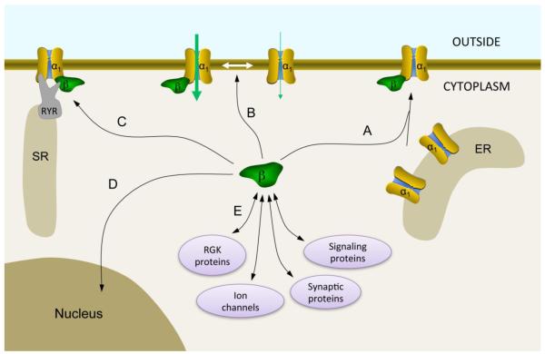
The voltage-gated Ca²⁺ channel β subunit (Ca(v)β) is a cytosolic auxiliary subunit that plays an essential role in regulating the surface expression and gating properties of high-voltage activated (HVA) Ca²⁺ channels. It is also crucial for the modulation of HVA Ca²⁺ channels by G proteins, kinases, Ras-related RGK GTPases, and other proteins. There are indications that Ca(v)β may carry out Ca²⁺ channel-independent functions. Ca(v)β knockouts are either non-viable or result in a severe pathophysiology, and mutations in Ca(v)β have been implicated in disease. In this article, we review the structure and various biological functions of Ca(v)β, as well as recent advances. This article is part of a Special Issue entitled: Calcium channels.


Figures

Figure 1
Figure 1
VGCC topology and the structure of the Cavβ core in complex with the AID. (A) Schematic representation of the predicted transmembrane topology of the α1 subunit of VGCC. The AID, marked in red, is located on the I-II linker. ‘P’ indicates the pore loops, located between transmembrane regions S5 and S6. ‘+’ indicates the charged amino acids in S4 – the voltage sensor. (B) Amino acid sequence alignment of the AID from the indicated calcium channel α1 subunits. Residues involved in interactions with Cavβ are marked in red, with the most critical residues underlined. Residue numbers are indicated on both sides of the sequence. (C) Cavβ is organized into 5 regions represented schematically in the upper panel. The lower panel shows the crystal structure of the Cavβ3 core in complex with the AID (PDB accession code 1VYT) with the following regions: N-terminus (light blue), the SH3 domain (gold), part of the HOOK region (purple, residues 121-169), and the GK domain (green). Residues 137-166 of the HOOK region were disordered and are not included. The AID region of Cav1.2 (residues 422 to 446) is colored in orange.
Figure 2
Figure 2
Major functions of Cavβ. (A) Cavβ enhances Cavα1 localization to the plasma membrane by preventing Cavα1 degradation and exposing ER export signals on Cavα1. (B) Cavβ promotes VGCC gating, resulting in an overall enhancement of current. (C) Cavβ interacts with the ryanodine receptor (RYR) in the sarcoplasmic reticulum (SR) of muscle cells and is critical for excitation-contraction coupling. (D) Cavβ can be translocated into the nucleus where it may participate in transcriptional regulation. (E) Cavβ interacts directly with many intracellular proteins that regulate VGCC function. The strongest of those regulators are RGK proteins, which potently inhibit VGCCs (reviewed in this issue by Colecraft and colleagues, also see [7]). Other partners include ion channels (e.g., BKCa and bestrophin), synaptic proteins (e.g., synaptotagmin I and RIMI), and signaling proteins (such as kinases, phosphatases, dynamin, and Ahnak) [for extensive review see 7].
Figure 3
Figure 3
Modulation of HVA Ca2+ channel gating by Cavβ. (A) Activation: Cavβ shifts the current-voltage curve (left panel) and the activation curve (right panel) to more hyperpolarized voltages. (B) Inactivation: Cavβ shifts the voltage dependence of inactivation to more hyperpolarized voltages, except β2a, which shifts it to more depolarized voltages (left panel). All Cavβ subunits speed the kinetics of inactivation, except β2a, which slows the kinetics of channel inactivation (right panel). All traces are schematic representations.

References

    1. Lacerda AE, Kim HS, Ruth P, Perez-Reyes E, Flockerzi V, Hofmann F, Birnbaumer L, Brown AM. Normalization of current kinetics by interaction between the alpha 1 and beta subunits of the skeletal muscle dihydropyridine-sensitive Ca2+ channel. Nature. 1991;352:527–530. - PubMed
    1. Curtis BM, Catterall WA. Purification of the calcium antagonist receptor of the voltage-sensitive calcium channel from skeletal muscle transverse tubules. Biochemistry. 1984;23:2113–2118. - PubMed
    1. Ruth P, Rohrkasten A, Biel M, Bosse E, Regulla S, Meyer HE, Flockerzi V, Hofmann F. Primary structure of the beta subunit of the DHP-sensitive calcium channel from skeletal muscle. Science. 1989;245:1115–1118. - PubMed
    1. Pragnell M, De Waard M, Mori Y, Tanabe T, Snutch TP, Campbell KP. Calcium channel beta-subunit binds to a conserved motif in the I-II cytoplasmic linker of the alpha 1-subunit. Nature. 1994;368:67–70. - PubMed
    1. Waard M. De, Witcher DR, Pragnell M, Liu H, Campbell KP. Properties of the alpha 1-beta anchoring site in voltage-dependent Ca2+ channels. J Biol Chem. 1995;270:12056–12064. - PubMed

Publication types

LinkOut - more resources