Review
. 2013 Jul-Aug:104-105:122-9.
doi: 10.1016/j.prostaglandins.2012.07.003. Epub 2012 Sep 3.

An emerging link in stem cell mobilization between activation of the complement cascade and the chemotactic gradient of sphingosine-1-phosphate

Affiliations
Review

An emerging link in stem cell mobilization between activation of the complement cascade and the chemotactic gradient of sphingosine-1-phosphate

Mariusz Z Ratajczak et al. Prostaglandins Other Lipid Mediat. 2013 Jul-Aug.

Abstract

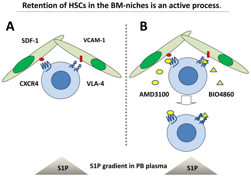
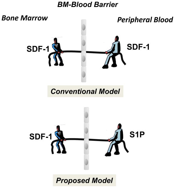
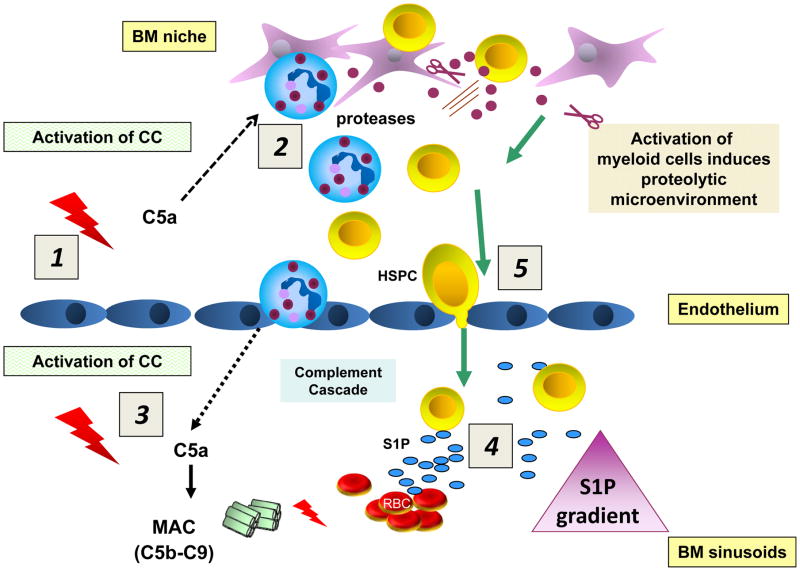
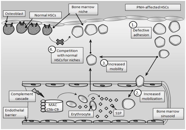
Under steady-state conditions, hematopoietic stem/progenitor cells (HSPCs) egress from bone marrow (BM) and enter peripheral blood (PB) where they circulate at low levels. Their number in PB, however, increases significantly in several stress situations related to infection, organ/tissue damage, or strenuous exercise. Pharmacologically mediated enforced egress of HSPCs from the BM microenvironment into PB is called "mobilization", and this phenomenon has been exploited in hematological transplantology as a means to obtain HSPCs for hematopoietic reconstitution. In this review we will present the accumulated evidence that innate immunity, including the complement cascade and the granulocyte/monocyte lineage, and the PB plasma level of the bioactive lipid sphingosine-1-phosphate (S1P) together orchestrate this evolutionarily conserved mechanism that directs trafficking of HSPCs.


Figures

Figure 1
Figure 1. HSPCs are actively retained in BM niches against the S1P chemotactic gradient between PB and BM
Panel A - HSPCs residing in BM are retained in niches due to active interaction between ligands expressed in the BM microenvironment (SDF-1 and VCMA-1) and their corresponding receptors (CXCR4 and VLA-4) present on the surface of HSPCs. Panel B – Blockage of these retention axes by small-molecule inhibitors (AMD3100 and BIO4860, respectively) exposes HSPCs to the S1P chemotactic gradient between PB and BM and leads to their egress from BM.
Figure 2
Figure 2. The concept of a chemotactic tug-of-war gradient between BM and PB explains mobilization and homing of HSPCs
It has been postulated that an SDF-1 gradient between BM and PB regulates trafficking of HSPCs (homing vs. mobilization). Under steady-state conditions, this gradient across endothelial barrier in BM sinusoids should be in balance. New evidence indicates that, rather than propsoed in conventional model changes in the SDF-1 gradient across the BM–PB barrier (e.g., by an increase of the SDF-1 level in PB), high S1P concentrations in PB plasma provide an opposing chemotactic gradient for HSPCs. However, since HSPCs are retained in BM in hematopoietic niches, the first step in mobilization is their release from these niches and permeabilization of endothelial barrier.
Figure 3
Figure 3. Activation of the CC directs mobilization of HSPCs
HSPCs are actively retained in BM and retention signals in the BM niches counteract an S1P-mediated chemotactic plasma gradient. Activation of the CC in the BM microenvironment (1) leads to generation of C5a, which strongly activates granulocytes and monocytes to release proteolytic enzymes that perturb retention signals for HSPCs in their niches (2). Activation of the CC in BM sinusoids also leads to release of C5a, which chemoattracts granulocytes and monocytes into PB (3). These cells are highly enriched in proteolytic enzymes and are the first to leave the BM and thus “pave the way” for HSPCs that follow in their “footsteps”. At the same time, activation of the CC in the BM sinusoids leads to generation of sublytic and lytic C5b-C9 (MAC), which promotes additional release of S1P from erythrocytes (4). In the final step, HSPCs egress from BM following the higher plasma S1P level in the BM sinusoids (5).
Figure 4
Figure 4. The role of CC activation and S1P-induced hypermobility of PNH-affected HSPCs
Due to a GPI-A-mediated defect in lipid raft formation, PNH HSPCs have impaired adhesion in BM niches (1), resulting in their enhanced egress (mobilization) from the BM into PB in response to an S1P gradient (2) and enhanced mobility in the BM microenvironment (3). Based on this finding, PNH HSPCs, which show higher motility than their normal healthy counterparts, outcompete and outgrow normal HSPCs in the BM microenvironment over time (4). These cells when complement cascade becomes activated and C5a - that permeabilizes blood-BM barrier is released, are preferentially as we demonstrated mobilized into PB [38]. Eventually, this defect in adhesion of PNH HSPCs in the BM microenvironment and their defective interaction with cellular and extracellular elements in the hematopoietic niche may trigger malignant transformation of HSPCs.

References

    1. Mikkola HKA, Orkin SH. The journey of developing hematopoietic stem cells. Development. 2006;133:3733–44. - PubMed
    1. Papayannopoulou T. Current mechanistic scenarios in hematopoietic stem/progenitor cell mobilization. Blood. 2004;103:1580–85. - PubMed
    1. Ratajczak MZ, Kim CH, Wojakowski W, Janowska-Wieczorek A, Kucia M, et al. Innate immunity as orchestrator of stem cell mobilization. Leukemia. 2010;24:1667–75. - PubMed
    1. Lévesque JP, Helwani FM, Winkler IG. The endosteal ‘osteoblastic’ niche and its role in hematopoietic stem cell homing and mobilization. Leukemia. 2010;24:1979–92. - PubMed
    1. Doan PL, Chute JP. The vascular niche: home for normal and malignant hematopoietic stem cells. Leukemia. 2012;26:54–62. - PubMed

Publication types

MeSH terms