

Magnetic transfer contrast accurately localizes substantia nigra confirmed by histology
- PMID: 22981657
- PMCID: PMC3534824
- DOI: 10.1016/j.biopsych.2012.07.035
Magnetic transfer contrast accurately localizes substantia nigra confirmed by histology
Abstract
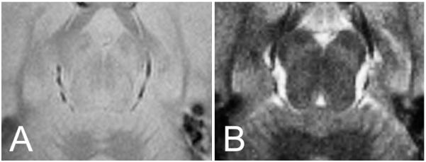
Background: Magnetic resonance imaging (MRI) has multiple contrast mechanisms. Like various staining techniques in histology, each contrast type reveals different information about the structure of the brain. However, it is not always clear how structures visible in MRI correspond to structures previously identified by histology. The purpose of this study was to determine if magnetic transfer contrast (MTC) or T2 contrast MRI was better at delineating the substantia nigra (SN).
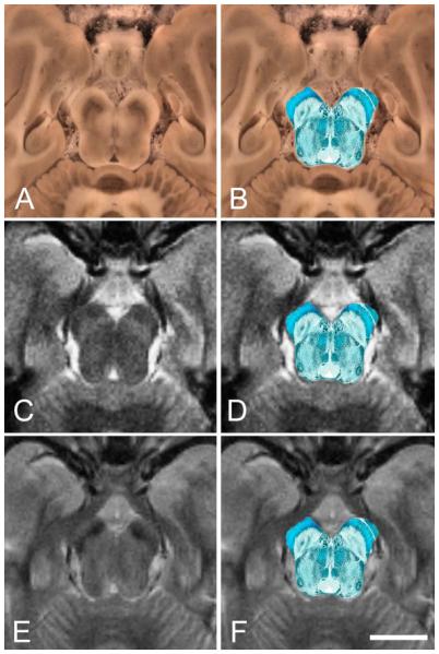
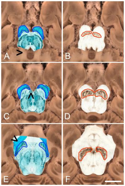
Methods: MRI scans were acquired in vivo from two nonhuman primates (NHPs). The NHPs were subsequently euthanized, perfused, and their brains sectioned for histologic analyses. Each slice was photographed before sectioning. Each brain was sectioned into approximately 500 sections, 40 μm each, encompassing most of the cortex, midbrain, and dorsal parts of the hindbrain. Levels corresponding to anatomic MRI images were selected. From these, adjacent sections were stained using Kluver-Barrera (myelin and cell bodies) or tyrosine hydroxylase (dopaminergic neurons) immunohistochemistry. The resulting images were coregistered to the block-face images using a moving least squares algorithm with similarity transformations. MR images were similarly coregistered to the block-face images, allowing the structures on MRI to be identified with structures on the histologic images.
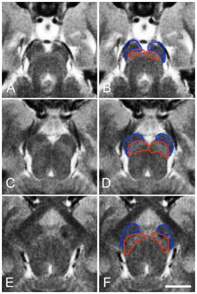
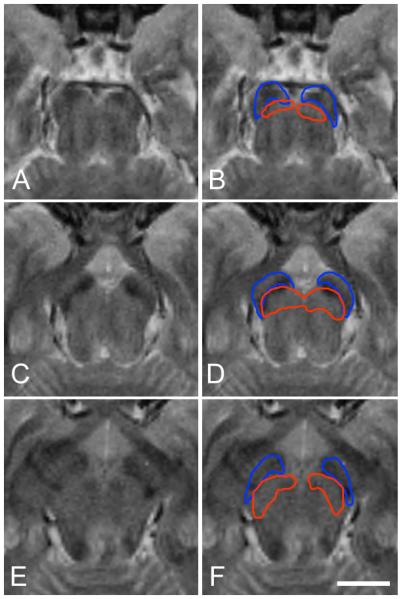
Results: We found that hyperintense (light) areas in MTC images were coextensive with the SN as delineated histologically. The hypointense (dark) areas in T2-weighted images were not coextensive with the SN but extended partially into the SN and partially into the cerebral peduncles.
Conclusions: MTC is more accurate than T2-weighting for localizing the SN in vivo.
Copyright © 2013 Society of Biological Psychiatry. Published by Elsevier Inc. All rights reserved.
Figures
References
-
- Shibata E, Sasaki M, Tohyama K, Otsuka K, Endoh J, Terayama Y, et al. Use of neuromelanin-sensitive MRI to distinguish schizophrenic and depressive patients and healthy individuals based on signal alterations in the substantia nigra and locus ceruleus. Biological psychiatry. 2008;64:401–406. - PubMed
-
- Menke RA, Scholz J, Miller KL, Deoni S, Jbabdi S, Matthews PM, et al. MRI characteristics of the substantia nigra in Parkinson’s disease: a combined quantitative T1 and DTI study. Neuroimage. 2009;47:435–441. - PubMed
-
- Enochs WS, Petherick P, Bogdanova A, Mohr U, Weissleder R. Paramagnetic metal scavenging by melanin: MR imaging. Radiology. 1997;204:417–423. - PubMed
Publication types
MeSH terms
Grants and funding
LinkOut - more resources
Full Text Sources
Other Literature Sources
Medical
