. 2012 Nov;130(5):1187-1196.e5.
doi: 10.1016/j.jaci.2012.07.031. Epub 2012 Sep 13.

Thymic stromal lymphopoietin is induced by respiratory syncytial virus-infected airway epithelial cells and promotes a type 2 response to infection

Affiliations

Thymic stromal lymphopoietin is induced by respiratory syncytial virus-infected airway epithelial cells and promotes a type 2 response to infection

Hai-Chon Lee et al. J Allergy Clin Immunol. 2012 Nov.

Abstract

Background: Respiratory viral infection, including respiratory syncytial virus (RSV) and rhinovirus, has been linked to respiratory disease in pediatric patients, including severe acute bronchiolitis and asthma exacerbation.

Objective: The study examined the role of the epithelial-derived cytokine thymic stromal lymphopoietin (TSLP) in the response to RSV infection.

Methods: Infection of human airway epithelial cells was used to examine TSLP induction after RSV infection. Air-liquid interface cultures from healthy children and children with asthma were also tested for TSLP production after infection. Finally, a mouse model was used to directly test the role of TSLP signaling in the response to RSV infection.

Results: Infection of airway epithelial cells with RSV led to the production of TSLP via activation of an innate signaling pathway that involved retinoic acid induced gene I, interferon promoter-stimulating factor 1, and nuclear factor-κB. Consistent with this observation, airway epithelial cells from asthmatic children a produced significantly greater levels of TSLP after RSV infection than cells from healthy children. In mouse models, RSV-induced TSLP expression was found to be critical for the development of immunopathology.

Conclusion: These findings suggest that RSV can use an innate antiviral signaling pathway to drive a potentially nonproductive immune response and has important implications for the role of TSLP in viral immune responses in general.


Conflict of interest statement

Disclosure of potential conflict of interest: The rest of the authors declare that they have no relevant conflicts of interest.

Figures

FIG 1
FIG 1
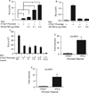
RSV induces TSLP expression in primary AECs. A, NHBECs were exposed to UV-irradiated RSV or RSV with indicated virus titers for 12 hours, and TSLP mRNA was measured by real-time quantitative PCR for virus titer. B and C, NHBECs were exposed to UV-irradiated RSV or RSV (MOI, 1) for indicated time course. Cell or culture supernatant fluids were harvested, and TSLP mRNA was measured by real-time quantitative PCR (Fig 1, B). TSLP protein levels were measured by ELISA (Fig 1, C). D and E, NHBECs were exposed to RSV (MOI, 1) without or with anti-TNFα antibodies (10, 50, or 100 ng/mL) (Fig 1, E), anti–IL-1R (1, 10, or 50 µg/mL) (Fig 1, E) for 24 hours, and culture supernatant fluids were harvested. TSLP protein levels were analyzed by ELISA. Data represent means ± SDs of 3 independent measurements. Similar results were obtained for 5 independent experiments (Fig 1, A–E). *P ≤ .05.
FIG 2
FIG 2
RIG-I and IPS-1 activation mediates Paramyxovirus-induced TSLP expression. A and B, A549 cells were transfected with control plasmid or luciferase constructs containing the human TSLP promoter alone or in combination with expression vectors for DN-RIG-I (Fig 2, A) or IPS-1 (Fig 2, B) followed by infection with RSV (MOI, 1). C, Normal littermate control (NLC) or RIG-I KO MEFs were infected with or without RSV, and TSLP protein expression was measured by ELISA. n.d. indicates not detected. D, NHBECs were transfected with control or RIG-I siRNA, followed by RSV infection, and TSLP mRNA was assessed at 8 hours after infection. Data represent means ± SDs of 3 independent measurements. Similar results were obtained for 5 independent experiments (Fig 2, A–D). *P ≤ .05.
FIG 3
FIG 3
RIG-I–mediated NF-κB activation controls SeV-induced TSLP expression. A and B, A549 cells were transfected with NF-κB p65 (Fig 3, A) and DN-IKKβ expression vectors with a human TSLP promoter construct (Fig 3, B). Cells were subsequently infected with SeV as above and analyzed for luciferase activity. Data are the means ± SDs with ≥3 for each group. C, A549 cells were transfected with control or constructs containing the WT hTSLP promoter or promoter NF-κB site mutants. WT, Nonmutated human TSLP promoter; 1, deletion of −3.2-kb site, 2, deletion of −1.3-kb site, 3, deletion of −0.2-kb site. Cells were subsequently infected with SeV as above and analyzed for luciferase activity. D and E, Human TSLP or IFNβ promoter reporters were cotransfected with expression vectors encoding IRF-3 (Fig 3, D) or IRF-7 (Fig 3, E) and infected with SeV. After 6 hours cell lysates were analyzed for luciferase activity. Data are the means ± SDs with ≥3 for each group. *P ≤ .05.
FIG 4
FIG 4
Human asthmatic epithelium produces greater levels of TSLP in response to RSV infection. ALI cultures were generated from primary BECs isolated from healthy or asthmatic children via bronchial brushing. Data show TSLP protein levels, measured by ELISA, in basolateral culture supernatant fluids after infection with either RSV Line 19 or RSV A2 or control at an MOI of 0.5 (n = 12 patients for all groups). P values were calculated with an ANOVA with Tukey posttest.
FIG 5
FIG 5
RSV-mediated expression of RIG-I and TLR3 in ALI cultures from healthy and asthmatic children. RIG-I and TLR3 expression (log2 scale) by BECs from healthy (open plots; n = 9) and asthmatic children (solid plots; n = 12) in response to RSV infection by A2 and Line 19 strains or exposure to control vero cell supernatant fluid (VC). RIG-I expression: ANOVA P < .001 between groups; asthma RSV A2 group 3-fold greater RIG-I expression than asthma VC group (*P < .001); asthma RSV 19 group 4-fold greater RIG-I expression than asthma VC group (*P < .001); no significant change in RIG-I expression by healthy cells with RSV A2 or RSV 19; no significant difference in RIG-I expression between asthmatic and healthy VC groups. TLR3 expression: ANOVA P = .003 between groups; healthy RSV A2 group 1.4-fold greater TLR3 expression than by healthy VC group (**P = .05); healthy RSV 19 group 1.8-fold greater TLR3 expression than by healthy VC group (***P = .03); asthma RSV A2 group 1.5-fold greater TLR3 expression than by asthma VC group (†P = .002); asthma RSV 19 group 1.8-fold greater TLR3 expression than by asthma VC group (††P = .002); no significant difference in TLR3 expression between asthmatic and healthy VC groups. RIG-I and TLR3 expression normalized to GAPDH. Bottom and top of box plots represent 25th and 75th percentiles, respectively; dotted band represents the median and whiskers represent minimums and maximums.
FIG 6
FIG 6
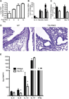
TSLP promotes RSV-mediated immunopathology in vivo. A, WT Balb/c mice were infected intratra-cheally with RSV and evaluated for TSLP expression in lung homogenate by ELISA at 6, 8, and 10 days after infection. B–D, WT or TSLPRKO mice were infected with RSV intratracheally and analyzed for immunopathology at day 6 after infection. B, IL-13 and mucin (Muc5AC, Gob5) mRNA expression between WT (filled bars) and TSLPRKO (open bars) mice. C, Airway resistance after methacholine challenge, measured by plethysmography, in RSV infected WT (filled bars) and TSLPRKO (open bars) mice infected with RSV for 8 days. D, PAS stain of lung section from WT or TSLPRKO mice after RSV infection. E, Cytokine production in supernatant fluids from restimulated mediastinal lymph node cultures from RSV-infected WT (filled bars) and TSLPRKO (open bars) mice. Data are representative of 3 experiments with 5 mice per group per experiment. *P ≤ .05.

Comment in

Similar articles

Cited by

References

    1. Sigurs N, Aljassim F, Kjellman B, Robinson PD, Sigurbergsson F, Bjarnason R, et al. Asthma and allergy patterns over 18 years after severe RSV bronchiolitis in the first year of life. Thorax. 2010;65:1045–1052. - PubMed
    1. Johnston SL, Pattemore PK, Sanderson G, Smith S, Lampe F, Josephs L, et al. Community study of role of viral infections in exacerbations of asthma in 9–11 year old children. BMJ. 1995;310:1225–1229. - PMC - PubMed
    1. Bueno SM, Gonzalez PA, Pacheco R, Leiva ED, Cautivo KM, Tobar HH, et al. Host immunity during RSV pathogenesis. Int Immunopharmacol. 2008;8:1320–1329. - PubMed
    1. Lukacs NW, Tekkanat KK, Berlin A, Hogaboam CM, Miller A, Evanoff HL, et al. Respiratory syncytial virus predisposes mice to augmented allergic airway responses via IL-13-mediated mechanisms. J Immunol. 2001;167:1060–1065. - PubMed
    1. Ying S, O’Connor B, Ratoff J, Meng Q, Mallett K, Cousins D, et al. Thymic stromal lymphopoietin expression is increased in asthmatic airways and correlates with expression of Th2-attracting chemokines and disease severity. J Immunol. 2005;174:8183–8190. - PubMed

Publication types

MeSH terms