. 2012 Dec;93(3):309-14.
doi: 10.1016/j.yexmp.2012.09.002. Epub 2012 Sep 12.

FAT10 knock out mice livers fail to develop Mallory-Denk bodies in the DDC mouse model

Affiliations

FAT10 knock out mice livers fail to develop Mallory-Denk bodies in the DDC mouse model

S W French et al. Exp Mol Pathol. 2012 Dec.

Abstract

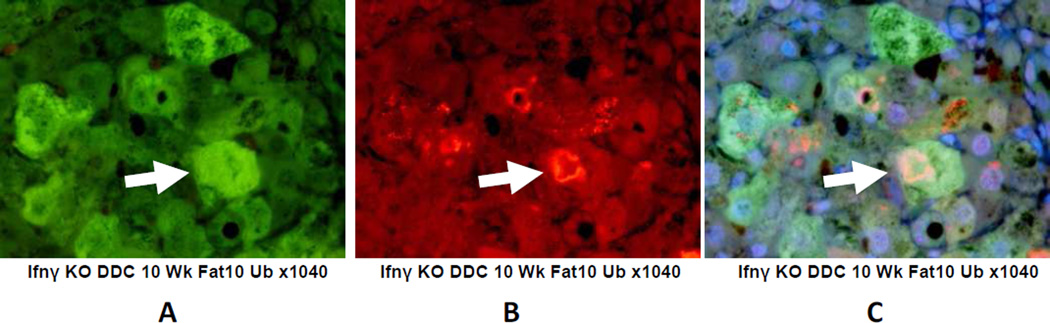
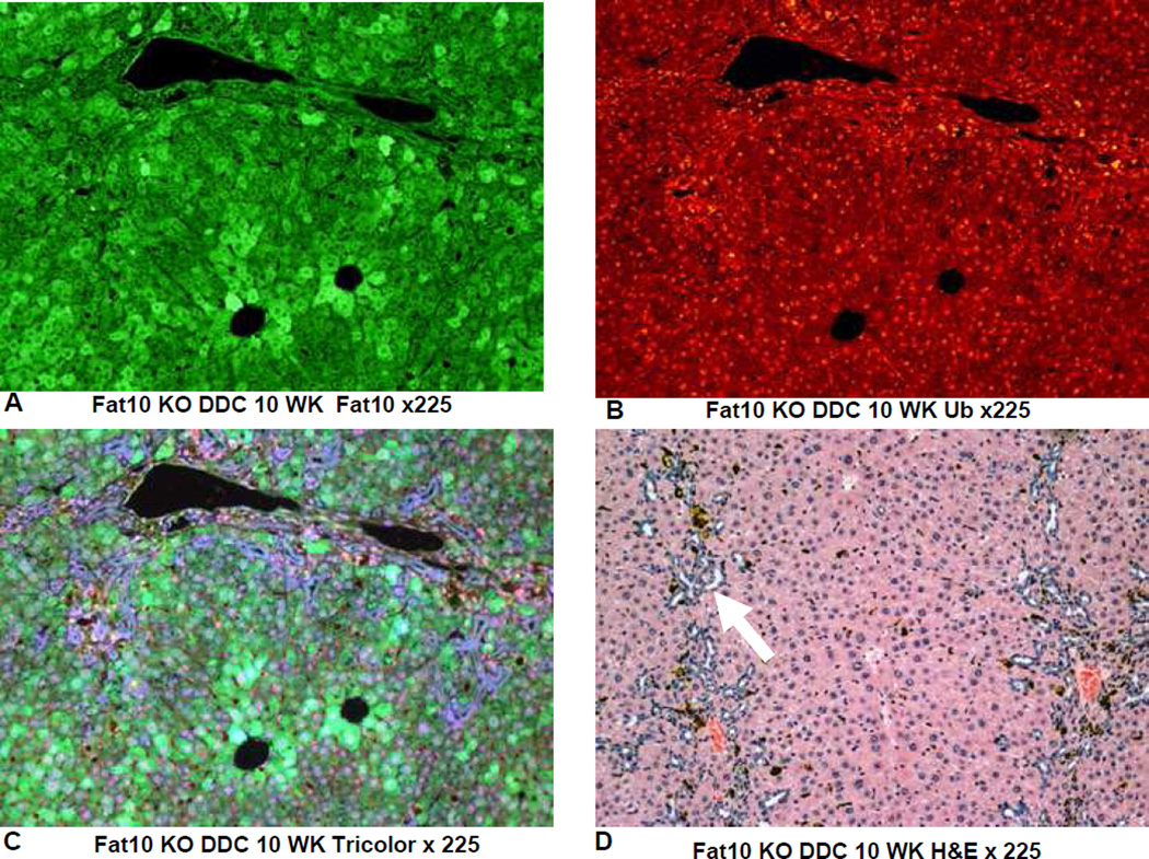
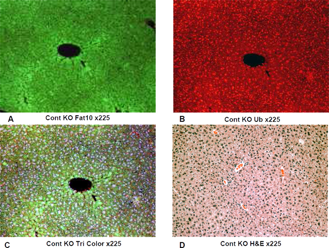
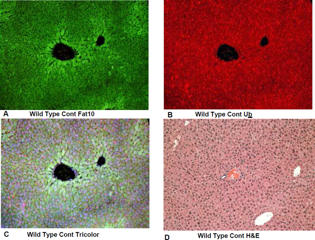
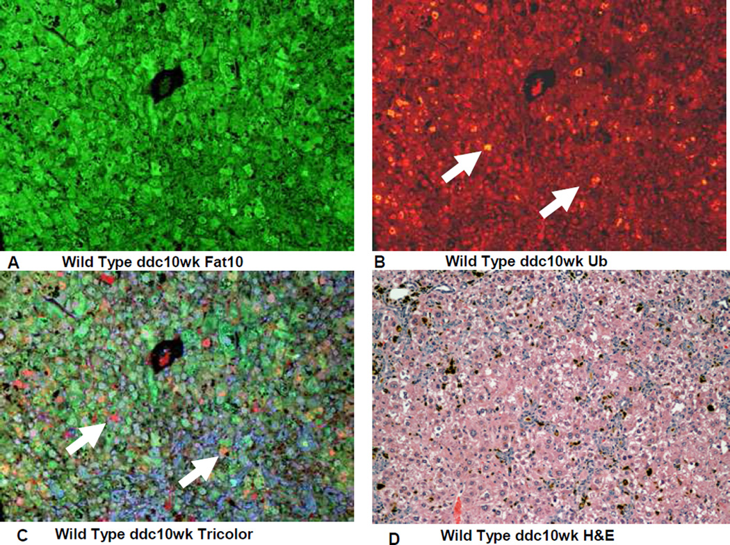
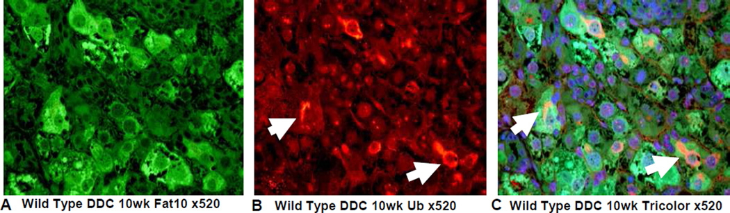
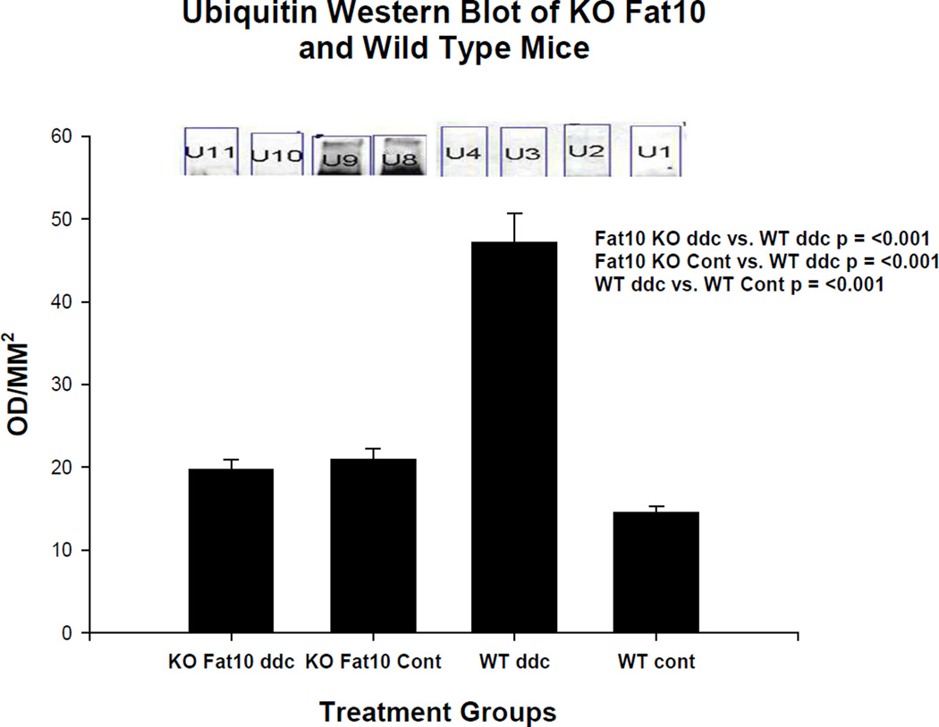
Mallory-Denk bodies (MDBs) are aggresomes composed of undigested ubiqutinated short lived proteins which have accumulated because of a decrease in the rate of their degradation by the 26s proteasome. The decrease in the activity of the proteasome is due to a shift in the activity of the 26s proteasome to the immunoproteasome triggered by an increase in expression of the catalytic subunits of the immunoproteasome which replaces the catalytic subunits of the 26s proteasome. This switch in the type of proteasome in liver cells is triggered by the binding of IFNγ to the IFNγ sequence response element (ISRE) located on the FAT10 promoter. To determine if either FAT10 or IFNγ are essential for the formation of MDBs we fed both IFNγ and FAT10 knock out (KO) mice DDC added to the control diet for 10weeks in order to induce MDBs. Mice fed the control diet and Wild type mice fed the DDC or control diet were compared. MDBs were located by immunofluorescent double stains using antibodies to ubiquitin to stain MDBs and FAT10 to localize the increased expression of FAT10 in MDB forming hepatocytes. We found that MDB formation occurred in the IFNγ KO mice but not in the FAT10 KO mice. Western blots showed an increase in the ubiquitin smears and decreases β 5 (chymotrypsin-like 26S proteasome subunit) in the Wild type mice fed DDC but not in the FAT10 KO mice fed DDC. To conclude, we have demonstrated that FAT10 is essential to the induction of MDB formation in the DDC fed mice.


Figures

Fig 1
Fig 1
Liver weights are shown comparing Interferon γ KO mice fed DDC for 10 weeks with Interferon γ wild type mice fed the diet without DDC added. The difference is statistically significant.
Fig 2
Fig 2
Liver weights are shown comparing four groups of mice. Group 1 was FAT10 KO mice fed DDC for 10 weeks, Group 2 was the FAT10 KO mice fed the diet without DDC added. Group 3 was wild type strain control mice fed DDC for 10 weeks. Group 4 was wild type strain control mice fed the diet without DDC added. Group 3 livers were significantly enlarged compared to the other 3 groups.
Fig 3
Fig 3
Immunohistochemical double stains (IHC) showing MDB formation in FAT10 positive hepatocytes. A) FAT10 antibody filter (FITC). B) Ubiquitin filter (Texas red) and C) tricolor filter showing MDB formed in FAT10 positive hepatocytes (arrows). X1040
Fig 4
Fig 4
The IHC double stains showing the absence of MDB formation in a liver from a FAT10 KO mouse fed DDC for 10 weeks. A) FAT10 antibody (FITC). B) Ubiquitin antibody filter (Texas red). C) Tricolor filter. D) The same liver stained with Hematoxylin and eosin showing proliferation of oval cells (arrow) induced by DDC feeding. X225
Fig 5
Fig 5
IHC double stains showing the FAT10 and ubiquitn of a liver from a FAT10 KO mouse fed the control diet without DDC added for 10 weeks. A) FAT10 antibody filter (FITC). B) Ubiquitin antibody filter (Texas red). C) Tricolor filter. D) The same liver stained with Hematoxylin and eosin showing normal liver histology. X225
Fig 6
Fig 6
IHC double stains showing the FAT10 and ubiquitin of a liver from a FAT10 wild type strain control mouse fed the control diet without DDC added. A) FAT10 antibody filter (FITC). B) Ubiquitin antibody filter (Texas red). C) Tricolor filter. D) The same liver stained with Hematoxylin and eosin showing normal liver histology. X225
Fig 7
Fig 7
IHC double stains showing the FAT10and ubiqutin of a liver from a FAT10 wild type strain control mouse fed the control diet with DDC added for 10 weeks. A) FAT10 antibody filter (FITC). B) Ubiquitin antibody filter (Texas red). C) Tricolor filter. Multiple ubiquitin positive MDBs are present (arrows) D) The same liver stained with Hematoxylin and eosin showing changed liver histology. X225
Fig 8
Fig 8
IHC double stains for FAT10 and ubiquitin of a liver from a wild type mouse fed DDC for 10 weeks show numerous MDBs formed by FAT10 positive liver cells (arrows). A) FAT10 antibody (Fitc). B) Ubiquitin antibody (Texas red). C) Tricolor filter. X520
Fig 9
Fig 9
Ubiquitin smear is present on a Western blot done on the wild type mouse fed DDC for 10 weeks (WT ddc) (U8 and U9). The other 3 groups of mice including the FAT10 KO mice fed DDC (KO FAT10 ddc) did not develop a ubiquitin smear indicating the activity of the 26s proteasome was not diminished in these groups of mice.
Fig 10
Fig 10
B5 (chymotrypsin) was diminished in the wild type mice fed DDC for 10 weeks (WT ddc) compared to the other 3 groups of mice by ½ including the FAT10 KO mice fed DDC (KO FAT ddc) indicating that the FAT10 KO mice fed DDC were prevented from developing a decrease in the 26s proteasome catalytic subunit because of the absence of FAT10 dependent decrease in the expression of Beta 5. The level of Beta 5 was slightly lower in the FAT10 KO DDC fed mice compared to the FAT10 KO controls and the WT controls.

Similar articles

Cited by

References

    1. Bardag-Gorce F, Oliva J, French BA, Li J, Dedes J, French SW. SAMe prevents the induction of immunoproteasome and preserves the 26s proteasomes in the DDC induced MDB; Mouse Model. Exp Mol Pathol. 2010a;88:352–362. - PMC - PubMed
    1. Bardag-Gorce F, Oliva J, Lin A, Li J, French BA, French SW. SAMe prevents the up regulation of toll-like receptor signaling in Mallory-Denk body forming hepatocytes. Exp Mol Pathol. 2010b;88:376–379. - PMC - PubMed
    1. Bardag-Gorce F, Oliva J, Villegas J, Fraley S, Amidi F, Li J, Dedes J, French B, French SW. Epigenetic mechanisms regulate Mallory Denk body formation in the livers of drug-primed mice. Exp Mol Pathol. 2008;94:113–121. - PMC - PubMed
    1. Bradford HM. A rapid and sensitive method for the quantification of microgram quantities of protein utilizing the principle of protein-dye binding. Anal Biochem. 1976;72:248–254. - PubMed
    1. Canaan A, Yu X, Booth CJ, Lian I, Lazar I, Gamfi SL, Castille K, Kohya N, Nakayama Y, Liu Y-C, Eynon E, Flavell R, Weissman SM. FAT10/diubiquitin-like protein-deficient mice exhibit minimal phenotypic differences. Mol Cell Biol. 2006;26:5180–5189. - PMC - PubMed

Publication types

MeSH terms