

Review
doi: 10.1016/j.sbi.2012.07.016.
Epub 2012 Sep 13.
Elements of ribosomal drug resistance and specificity
Affiliations
- PMID: 22981944
- PMCID: PMC5560274
- DOI: 10.1016/j.sbi.2012.07.016
Item in Clipboard
Review
Elements of ribosomal drug resistance and specificity
Curr Opin Struct Biol.
2012 Dec.
Abstract
The structures of ribosomes in complex with inhibitors of translation have not only shed light on the interactions of antibiotics with the ribosome but also on the underlying mechanisms by which they interfere with the ribosome function. Several recent papers [1(•),2(••),3,4] have correlated the available ribosome structures with the wealth of biochemical data [5(•)]. In this review we shall focus on the lessons learned for drug specificity rather than presenting a comprehensive survey of the known structures of ribosome complexes with antibiotics.
Copyright © 2012 Elsevier Ltd. All rights reserved.
Figures
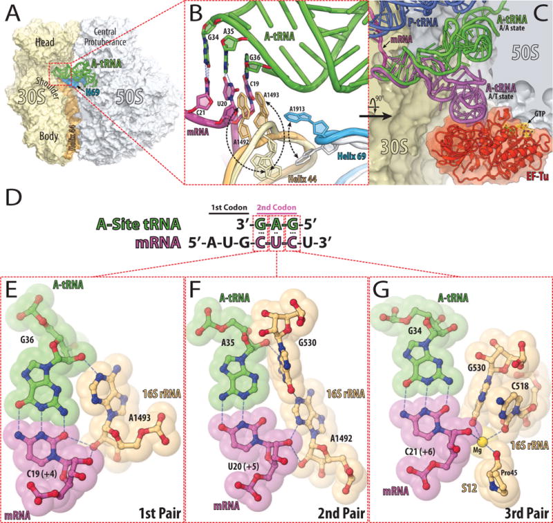
(A) The structure of 70S ribosome from eubacteria Thermus thermophilus with bound tRNAs and mRNA [57]. The 30S subunit is shown in light yellow with h44 of the 16S rRNA in light orange. The 50S subunit is in light blue with H69 of the 23S rRNA in marine. The tRNA bound in the A-site is in green. (B) Close up view of the decoding center of the 70S ribosome in the unliganded [58] and tRNA bound states [57]. The subunits and A-site bound tRNA are colored as in (A), the mRNA is in magenta. The conformational change of each nucleotide upon tRNA binding is indicated with black dashed arrows. Nucleotides involved in the codon-anticodon interactions (hydrogen bonds in blue dashed lines) are represented as sticks with their nitrogen and oxygen atoms in dark blue and red, respectively. (C) Comparison of the tRNA in the A/T state (purple) [59] with the accommodated A/A state (green) [57]. EF-Tu is shown in red, GTP in yellow, the 30S subunit in light yellow, 50S subunit in light blue, the P-site bound tRNA in dark blue, and the mRNA in magenta. (D) Schematic diagram of codon-anticodon interactions between mRNA (magenta) and cognate tRNA (green). (E, F, G) Codon-anticodon recognition by nucleotides of the 16S rRNA [60]. Cognate A-site bound tRNA is displayed in green, mRNA in magenta, nucleotides of 16S rRNA and portion of protein S12 are in light orange. Nitrogen, oxygen, and magnesium atoms are colored in blue, red, and yellow, respectively.
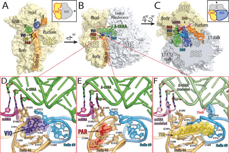
(A, B, C) Overview of the superimposed binding sites of viomycin (purple) [18], paromomycin (red) [57], and thermorubin (yellow) [26] on the Thermus thermophilus 70S ribosome viewed from three different perspectives. Shown in light yellow is the 30S subunit with h44 in light orange and in light blue is the 50S subunit with H69 in marine. The tRNAs are displayed in green for the A-site, in dark blue for the P-site, and in orange for the E-site bound tRNA. The mRNA is shown in magenta. In (A), the 30S subunit is viewed from the 50S subunit, as indicated by the inset. The view in (B) is from the cytoplasm onto the A-site. The view in (C) is from the top after removing the head of the 30S subunit and protuberances of the 50S subunit, as indicated by the inset. Close up views of the binding sites of viomycin (VIO, purple), paromomycin (PAR, red), and thermorubin (TER, yellow) are shown in panels (D), (E), and (F), respectively.
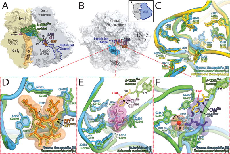
(A, B) Overview of the superimposed binding sites of erythromycin (ERY, orange), clindamycin (CLI, pink), and chloramphenicol (CAM, purple) bound to the Thermus thermophilus 70S ribosome viewed from two different perspectives. (A) 70S ribosome is cut open along the exit tunnel. Shown in light yellow is the 30S subunit with h44 in light orange and in light blue is the 50S subunit with H69 in marine. The mRNA is shown in magenta. Bound to the A-site is an amino-acylated tRNAPhe shown in green with its 3′-terminal phenylalanine residue in yellow. (B) The 50S subunit viewed from the 30S subunit as indicated by the inset and with the same color-coding as in (A). (C) A close up view of the superimposed antibiotic binding sites in ribosomes from the three different kingdoms of life. The drugs are omitted for clarity. As an example of the eubacterial binding site (B) the 23S rRNA from Thermus thermophilus (blue) is shown, for arecheal (A) the 23S rRNA from Haloarcula marismortui (green), and for eukaryotic (E) the 28S rRNA from Tetrahymena thermophila (yellow). (D, E, F) Close up views of the comparisons between eubacterial (blue) and archaeal (green) antibiotic binding sites. (D) Comparison of the structures of erythromycin (ERY) bound to Thermus thermophilus 70S ribosome (orange) and bound to Haloarcula marismortui 50S subunit carrying a G2099A mutation (yellow). (E) Clindamycin bound to Escherichia coli 70S ribosome is displayed in dark pink and in light pink when bound to Haloarcula marismortui 50S subunit carrying a G2099A mutation. (F) Chloramphenicol bound to Thermus thermophilus 70S ribosome is displayed in purple and in light blue is anisomycin when bound to Haloarcula marismortui 50S subunit. Potassium ion is shown in red.
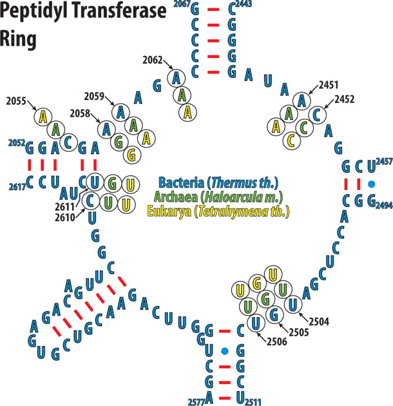
Nucleotides of the peptidyl transferase ring of the eubacteria Thermus thermophilus are shown in blue. The phylogenetic variations of the nucleotides discussed in the text are shown in green for archeon Haloarcula marismortui, and yellow for eukaryote Tetrahymena thermophila.
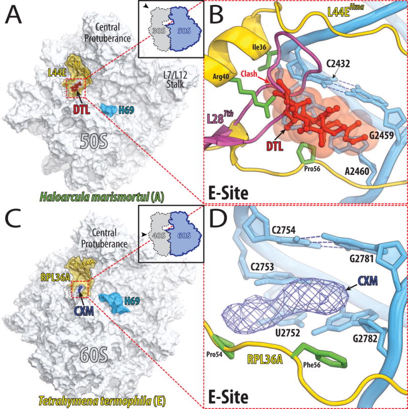
(A) Overview of the binding sites of 13-deoxytedanolide (DTL, red) on the large ribosomal subunit from archaeon Haloarcula marismortui as viewed from the 30S subunit [50]. Black contour outlines L44e protein, which surrounds the E-site and whose visible portion is highlighted in yellow. (B) A close up view of the 13-deoxytedanolide binding site. The 23S rRNA is colored in blue and L44e is in yellow with its Ile36, Arg40, and Pro56 side-chains in green. A model of the L28 ribosomal protein from Thermus thermophilus is shown in pink. Note, that L28 partially occupies the space required for 13-deoxytedanolide binding to ribosome. (C) Overview of the cycloheximide binding site on the 60S ribosomal subunit from eukaryote Tetrahymena thermophila [53]. The 60S subunit (light blue) is viewed from the 40S ribosomal subunit. Colored in yellow is the visible portion of RPL36A, which encircles the E-site. The electron density map for cycloheximide (CXM) contoured at 3σ is represented as a blue mesh. (D) A close up view of the cycloheximide binding site. The 26S rRNA is colored in blue and RPL36A is in yellow with the Pro54 and Phe56 side-chains in green. The electron density map for cycloheximide (CXM) is represented as a blue mesh.
References
-
- Wilson DN. The A–Z of bacterial translation inhibitors. Critical Reviews in Biochemistry and Molecular Biology. 2009;44:393–433. An extensive review of the recent structures of inhibitors bound to the ribosome. - PubMed
-
- Sohmen D, Harms JM, Schlunzen F, Wilson DN. Enhanced SnapShot: Antibiotic inhibition of protein synthesis II. Cell. 2009;139:212–212 e211. An appealing graphical representation of the effects of many different classes of inhibitors on protein translation. - PubMed
-
- Poehlsgaard J, Douthwaite S. The bacterial ribosome as a target for antibiotics. Nat Rev Microbiol. 2005;3:870–881. - PubMed
-
- Kannan K, Mankin AS. Macrolide antibiotics in the ribosome exit tunnel: species-specific binding and action. Antimicrobial Therapeutics Reviews: Antibiotics That Target the Ribosome. 2011;1241:33–47. - PubMed
-
- Spahn CM, Prescott CD. Throwing a spanner in the works: antibiotics and the translation apparatus. J Mol Med (Berl) 1996;74:423–439. in combination with [1] and [41] represent most of the knowledge accumulated on inhibitors of protein translation over the past decades. - PubMed
Publication types
MeSH terms
Substances
Grants and funding
LinkOut - more resources
Full Text Sources
Medical
