Review
. 2013 Jan:32:102-80.
doi: 10.1016/j.preteyeres.2012.08.004. Epub 2012 Sep 11.

Role of the retinal vascular endothelial cell in ocular disease

Affiliations
Review

Role of the retinal vascular endothelial cell in ocular disease

Arpita S Bharadwaj et al. Prog Retin Eye Res. 2013 Jan.

Abstract

Retinal endothelial cells line the arborizing microvasculature that supplies and drains the neural retina. The anatomical and physiological characteristics of these endothelial cells are consistent with nutritional requirements and protection of a tissue critical to vision. On the one hand, the endothelium must ensure the supply of oxygen and other nutrients to the metabolically active retina, and allow access to circulating cells that maintain the vasculature or survey the retina for the presence of potential pathogens. On the other hand, the endothelium contributes to the blood-retinal barrier that protects the retina by excluding circulating molecular toxins, microorganisms, and pro-inflammatory leukocytes. Features required to fulfill these functions may also predispose to disease processes, such as retinal vascular leakage and neovascularization, and trafficking of microbes and inflammatory cells. Thus, the retinal endothelial cell is a key participant in retinal ischemic vasculopathies that include diabetic retinopathy and retinopathy of prematurity, and retinal inflammation or infection, as occurs in posterior uveitis. Using gene expression and proteomic profiling, it has been possible to explore the molecular phenotype of the human retinal endothelial cell and contribute to understanding of the pathogenesis of these diseases. In addition to providing support for the involvement of well-characterized endothelial molecules, profiling has the power to identify new players in retinal pathologies. Findings may have implications for the design of new biological therapies. Additional progress in this field is anticipated as other technologies, including epigenetic profiling methods, whole transcriptome shotgun sequencing, and metabolomics, are used to study the human retinal endothelial cell.


Figures

Figure 1
Figure 1
(A) Multi-dimensional scaling plot shows global gene expression by retinal endothelial cells from 6 human donors. Circles designate individual donors. (B) Relative gene expression of selected adhesion molecules in retinal endothelial cells from the same human donors. Normalized fluorescence intensity, which reflects hybridization to the relevant array probes, was averaged for each donor and expressed in log2 scale. Generated using previously published data (Smith et al., 2007).
Figure 1
Figure 1
(A) Multi-dimensional scaling plot shows global gene expression by retinal endothelial cells from 6 human donors. Circles designate individual donors. (B) Relative gene expression of selected adhesion molecules in retinal endothelial cells from the same human donors. Normalized fluorescence intensity, which reflects hybridization to the relevant array probes, was averaged for each donor and expressed in log2 scale. Generated using previously published data (Smith et al., 2007).
Figure 2
Figure 2
Photomicrographs of human retinal endothelial cells immortalized by transduction with LXSN16E6E7. Cell retain an endothelial phenotype as indicated by: (A) Cobblestone morphology. Original magnification: 200X; (B) Capillary-like tube formation after 24-hour incubation in 5% CO2 and at 37 °C on Matrigel (BD Biosciences Discovery Labware, Bedford, MA). Original Magnification: 100X. (C–D) Expression of (C) CD31, as detected by mouse monoclonal anti-human CD31 antibody (concentration: 10 μg/ml; clone: JC70A: isotype IgG1κ; BD Pharmingen Biosciences, San Diego, CA) and Alexa Fluor 488-conjugated goat anti-mouse IgG antibody (concentration: 5 μg/ml; Life Technologies, Molecular Probes, Eugene, OR) and (D) VWF, as detected by Alexa Fluor 488-conjugated rabbit polyclonal anti-human VWF antibody (concentration: 16 μg/ml; fraction: IgG; DAKO, Glostrup, Denmark) and Alexa Fluor 488-conjugated anti-rabbit IgG antibody (concentration: 5 μg/ml; Life Technologies, Molecular Probes). Propidium iodide nuclear counterstain (Life Technologies, Molecular Probes). Original magnification: 400X. Paired cell cultures stained with antibody directed against an irrelevant antigen showed no positive staining.
Figure 3
Figure 3
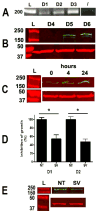
Gel images of supervillin (A) RT-PCR product (165 bp) and (B) protein (205 kDa, β-actin at 42 kDa) from primary retinal endothelial cells of 6 human donors (D1–D6). L = ladder./ = no cDNA. (C) Gel image of supervillin protein in human retinal endothelial cells stimulated with VEGF (20 ng/ml, Millipore, Temecula, CA) for 0, 4 and 24 hours. (D) Graph showing proliferation of immortalized retinal endothelial cells from 2 human donors initially plated at 3000 cells/wells in a 96-well plate, 96 hours after transfection with supervillin (SV) or non-targeted (NT) siRNA using Targefect-siRNA transfection kit (Targeting Systems, El Cajon, CA). SV siRNA: sense = 5′-GGCGGUCCCUCAUCAAGAAGC-3′, anti-sense = 5′-UUCUUGAUGAGGGACCGCCCU-3′ (designed using siDirect (Naito et al., 2004)). NT siRNA: sense = 5′-CGCCGACGUUUAACGGAAGCC-3′, anti-sense = 5′-CUUCCGUUAAACGUCGGCGCA-3′. n = 4–8 wells/condition. D = donor. * = p ≤ 0.003. (E) Gel image of supervillin protein in the siRNA-treated human retinal endothelial cells that were used in the proliferation experiment shown in (D – donor 1), 48 hours following transfection.
Figure 4
Figure 4
Graphs showing relative expression of E-selectin, P-selectin, ICAM-1, VCAM-1, and CD44 transcript by immortalized human retinal endothelial cells following exposure to one of the following conditions: medium alone; TNF-α (10ng/ml, R&D Systems, Minneapolis, MN); IFN-γ (20ng/ml, R&D Systems); and IL-17 (100ng/ml, R&D Systems). Endothelial cells were cultured to confluence in modified MCDB-131 medium (Sigma-Aldrich, St. Louis, MO) with 2.5% FBS (Hyclone, Logan, UT) and endothelial growth factors (EGM-2 SingleQuots supplement (Clonetics-Lonza, St. Louis, MO), omitting gentamicin, hydrocortisone and serum, at 1:4 dilution), and subsequently incubated with or without cytokine for 4 hours. Total RNA was isolated using the RNeasy Mini Kit (Qiagen, Valencia, CA), and cDNA was synthesized using the iScript cDNA Synthesis Kit (Bio-Rad Laboratories, Hercules, CA). Relative expression of gene products, normalized to GAPDH, was determined by using the Chromo4 Thermocycler and iQ SYBR Green Supermix (both from Bio-Rad Laboratories). Data were analyzed using Chromo4 Opticon Monitor 3 software. Primer sequences appear in Table 2. In all graphs, bars represent mean and error bars represent standard error of mean (n = 3 wells; * = p<0.05, ** = p<0.01, two-tailed Student’s t-test).
Figure 5
Figure 5
Graphs showing relative expression of CXCL10 and CCL20 transcript by immortalized human retinal endothelial cells following exposure to one of the following conditions: medium alone; TNF-α (10ng/ml); IFN-γ (20ng/ml); and IL-17 (100ng/ml). Experimental conditions and real-time quantitative RT-PCR are described in the Figure 4 legend. Primer sequences appear in Table 2. In both graphs, bars represent mean and error bars represent standard error of mean (n = 3 wells; * = p<0.05, ** = p<0.01, two-tailed Student’s t-test).
Figure 6
Figure 6
Graphs showing increased relative expression of VEGF165 transcript by immortalized human retinal endothelial cells following a 48-hour exposure to hypoxia. Endothelial cells were cultured to confluence in modified MCDB-131 medium (Sigma-Aldrich, St. Louis, MO) with 2% FBS (Hyclone, Logan, UT) and endothelial growth factors (EGM-2 SingleQuots supplement (Clonetics-Lonza, St. Louis, MO), omitting gentamicin, hydrocortisone and serum), and subsequently incubated for 48 hours in 1% or room air level oxygen. Total RNA was isolated using the RNeasy Mini Kit (Qiagen, Valencia, CA), and cDNA was synthesized using the iScript cDNA Synthesis Kit (Bio-Rad Laboratories, Hercules, CA). Relative expression of gene products, normalized to GAPDH, was determined by using the Chromo4 Thermocycler and iQ SYBR Green Supermix (both from Bio-Rad Laboratories). Data were analyzed using Chromo4 Opticon Monitor 3 software. Primer sequences appear in Table 2. Bars represent mean and error bars represent standard error of mean (n = 3 reactions; * = p < 0.001, two-tailed Student’s t-test).
Figure 7
Figure 7
Photomicrographs of fresh frozen retinal sections from the same human donor immunostained with rabbit anti-human ephrin-B2 polyclonal antibody (1:50 dilution; Santa Cruz Biotechnology, Santa Cruz, CA) diluted 1:50 or rabbit IgG similarly diluted as negative control. Specific staining was identified by Alexa Fluor 594-labelled secondary antibody (1:400 dilution; Invitrogen). (A) Retinal vascular channel expressing ephrin-B2; (B) Absence of positive staining in negative control. Original Magnification: 400X; (C) Ephrin-B2-positive arterial channel (arrowhead) adjacent to ephrin-B2-negative venous channel (arrow).

References

    1. Adamis AP, Shima DT, Yeo KT, Yeo TK, Brown LF, Berse B, D’Amore PA, Folkman J. Synthesis and secretion of vascular permeability factor/vascular endothelial growth factor by human retinal pigment epithelial cells. Biochem Biophys Res Commun. 1993;193:631–638. - PubMed
    1. Aguilar E, Dorrell MI, Friedlander D, Jacobson RA, Johnson A, Marchetti V, Moreno SK, Ritter MR, Friedlander M. Chapter 6. Ocular models of angiogenesis. Methods Enzymol. 2008;444:115–158. - PubMed
    1. Ahn JK, Yu HG, Chung H, Park YG. Intraocular cytokine environment in active Behcet uveitis. American journal of ophthalmology. 2006;142:429–434. - PubMed
    1. Aiello LP, Avery RL, Arrigg PG, Keyt BA, Jampel HD, Shah ST, Pasquale LR, Thieme H, Iwamoto MA, Park JE, et al. Vascular endothelial growth factor in ocular fluid of patients with diabetic retinopathy and other retinal disorders. The New England journal of medicine. 1994;331:1480–1487. - PubMed
    1. Aird WC. Mechanisms of endothelial cell heterogeneity in health and disease. Circ Res. 2006;98:159–162. - PubMed

Publication types

MeSH terms