Comparative Study
. 2012 Dec;1823(12):2179-89.
doi: 10.1016/j.bbamcr.2012.08.021. Epub 2012 Sep 6.

Endoplasmic reticulum stress induces the expression of COX-2 through activation of eIF2α, p38-MAPK and NF-κB in advanced glycation end products stimulated human chondrocytes

Affiliations
Comparative Study

Endoplasmic reticulum stress induces the expression of COX-2 through activation of eIF2α, p38-MAPK and NF-κB in advanced glycation end products stimulated human chondrocytes

Zafar Rasheed et al. Biochim Biophys Acta. 2012 Dec.

Abstract

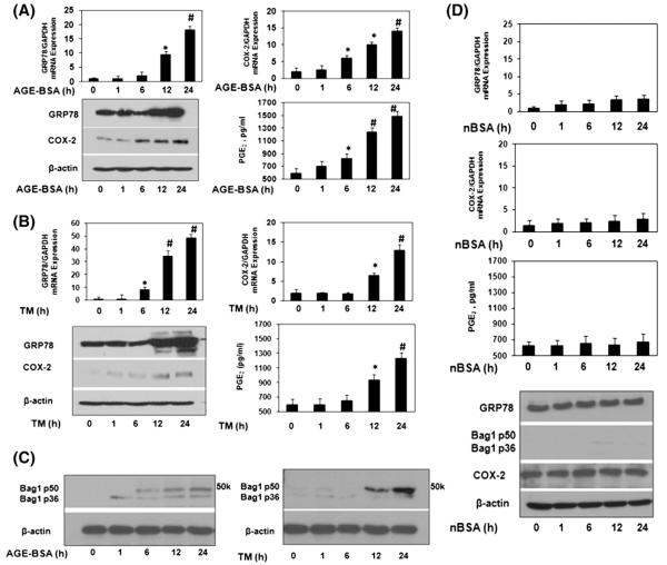
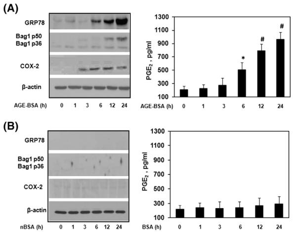
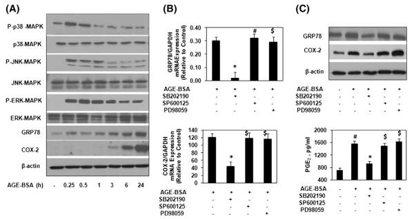
Introduction: During aging, advanced glycation end products (AGEs) accumulate in articular cartilage. In this study we determined whether AGEs induce endoplasmic reticulum (ER) stress and studied the ER stress-activated pathways that stimulate cyclooxygenase-2 (COX-2) expression in human chondrocytes.

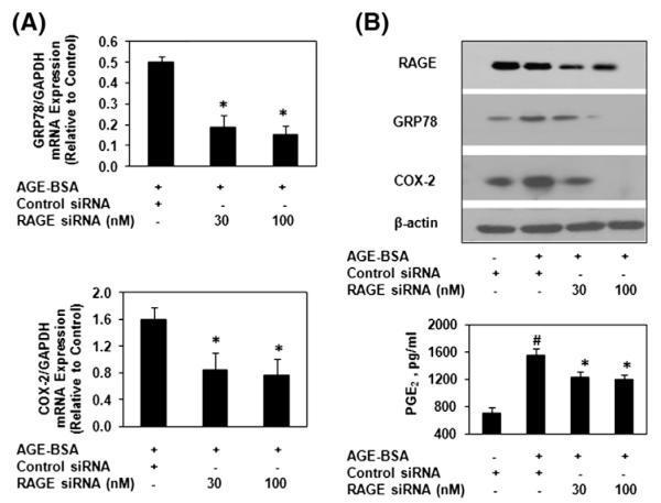
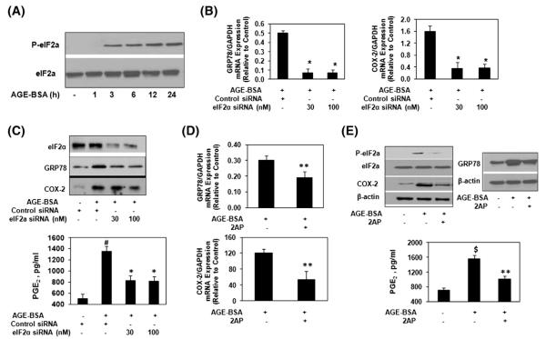
Methods: Chondrocytes were stimulated with AGE-BSA. Gene expression was determined by quantitative PCR and protein expression was studied by immunoblotting. Studies to elucidate involved pathways were executed using siRNAs and specific inhibitors of eukaryotic initiation factor-2α (eIF2α), MAPKs and NF-κB.

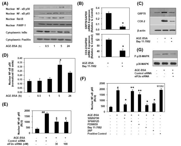
Results: AGE-BSA induced expression of GRP78 with concomitant increase in COX-2 expression was observed in human chondrocytes. In addition, expression of Bag-1, an ER stress marker was also increased by AGE-BSA. RAGE knockdown inhibited AGE-BSA-induced expression of GRP78 and COX-2. Treatment with eIF2α inhibitor or eIF2α knockdown inhibited AGE-BSA-induced expression of GRP78 and COX-2 with decreased PGE(2) production. Treatment with SB202190 inhibited AGE-BSA-induced expression of GRP78 and COX-2, while treatment with PD98051 inhibited AGE-BSA-induced GRP78 protein expression but had no effect on COX-2 protein expression. SP600125 had no effect on either GRP78 or COX-2 protein expression. Bay 11-7082 suppressed AGE-BSA-induced GRP78 and COX-2 expression. AGE-BSA-induced activation of NF-κB was inhibited by treatment with SB202190 and by eIF2α knockdown, but was not inhibited when chondrocytes were treated with SP600125 or PD98059.

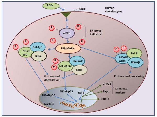
Conclusion: This study demonstrates that AGEs induce ER stress and stimulate the expression of COX-2 through eIF2α, p38-MAPK and NF-κB pathways in human chondrocytes. Our results provide important insights into cartilage degradation in osteoarthritis associated with latent ER stress.


Figures

Fig. 1
Fig. 1
Elevated expression of GRP-78, Bag-1 and COX-2 in AGE-BSA stimulated human chondrocytes. (A) Human OA chondrocytes (70–80% confluent) were treated with AGE-BSA (100 μg/ml) for 0, 1, 6, 12 and 24 h. Gene expression of GRP78 and COX-2 was determined by real time quantitative PCR and normalized to GAPDH and compared to the levels present in untreated OA chondrocytes using comparative ΔΔCT method. Cell lyastes were analyzed by immunoblotting with antibodies specific for GRP78 (cat. #3183, Cell Signaling Tech), COX-2 (cat. #4842, Cell Signaling Tech) and β-actin (cat. #sc-47778, Santa Cruz Biotech). β-actin was used as a loading control. Production of PGE2 in the culture medium was quantified by ELISA. (B) Human OA chondrocytes were treated with tunicamycin, TM (2.5 μM) for 0, 1, 6, 12 and 24 h. Gene or protein expressions were determined as described in section A. (C) Bag-1 expression in human OA chondrocytes stimulated by AGE-BSA or TM. Cell lyastes were analyzed by immunoblotting with antibodies specific for Bag-1 (cat. #sc-135844, Santa Cruz Biotech). (D) Human OA chondrocytes were treated with native (n)BSA (100 μg/ml) for 0, 1, 6, 12 and 24 h. Gene or protein expressions were determined as described in section A. Western blots are from single chondrocytes sample, but are representative of three chondrocytes samples. Bars show the mean with 95% confidence interval (CI) results of three independent assays, each of which run in triplicate experiments. *p<0.05 versus control; #p<0.0001 versus control.
Fig. 2
Fig. 2
Elevated expression of GRP78, Bag1 and COX-2 in AGE-BSA stimulated normal human chondrocytes. Normal human chondrocytes (70–80% confluent) were treated with AGE-BSA (100 μg/ml) (A) or with nBSA (100 μg/ml) (B) for 0, 1, 6, 12 and 24 h. Cell lyastes were analyzed by immunoblotting with antibodies specific for GRP78, COX-2. β-actin was used as a loading control. Production of PGE2 in the culture medium was quantified by ELISA. Normal chondrocytes were obtained from trauma patients. Immunoblots are from single chondrocytes sample, but are representative of two chondrocytes samples. Bars show the mean with 95% CI results of two independent assays, each of which run in triplicate experiments. *p<0.05 versus control; #p<0.0001 versus control.
Fig. 3
Fig. 3
AGE-BSA induced ER stress is mediated through RAGE in human chondrocytes. (A) RAGE-specific siRNA transfection on AGE-BSA-induced expression of GRP78 and COX-2 in OA chondrocytes. OA chondrocytes were transfected with RAGE-siRNA or control siRNA and then stimulated with AGE-BSA for 24 h. (A) Expression of GRP78 and COX-2 mRNA was determined as described under Fig. 1. (B) Protein expression of RAGE, GRP78 and COX-2 was determined by Immunoblot analysis. β-actin was used as a loading control. Production of PGE2 was quantified by ELISA. Immunoblots are from single chondrocytes sample, but are representative of five chondrocytes samples. Bars show the mean with 95% CI results of five independent assays, each of which run in triplicate experiments. #p<0.05 versus chondrocytes treated with AGE-BSA+control siRNA; *p<0.001 versus chondrocytes transfected with control siRNA alone.
Fig. 4
Fig. 4
AGE-BSA-induced stimulation of GRP78 and COX-2 is mediated through α subunit of eIF2 in human chondrocytes. (A) Human OA chondrocytes were treated with AGE-BSA for the indicated times, the phosphorylation of eIF2α was determined by immunobloting. (B) eIF2α-specific siRNA transfection and gene expression of GRP78 and COX-2 in AGE-BSA-stimulated OA chondrocytes. (C) eIF2α-specific siRNA transfection and protein expression of GRP78 and COX-2 in AGE-BSA-stimulated OA chondrocytes. (D) eIF2α-phosphorylation inhibitor 2AP and gene expression of GRP78 and COX-2. OA chondrocytes were pretreated with 2AP (10 μM) for 2 h and stimulated with AGE-BSA for 24 h. (E) 2AP and protein expression of eIF2α, GRP78 and COX-2 or PGE2 production. OA chondrocytes were pretreated with 2AP for 2 h and stimulated with AGE-BSA for 24 h. See Fig. 1 for details. Western immunoblots are from single chondrocytes sample, but are representative of three chondrocytes samples. Bars show the mean with 95% CI results of three independent experiments, each of which run in triplicate experiments. *p<0.001 versus chondrocytes treated with AGE-BSA+control siRNA; #p<0.05 versus chondrocytes transfected with control siRNA; **p<0.05 versus chondrocytes treated with AGE-BSA alone; $p<0.0001 versus untreated chondrocytes.
Fig. 5
Fig. 5
AGE-BSA-induced activation of p38-MAPKs is essential for COX-2 induction during ER stress in human chondrocytes. (A) Human OA chondrocytes were treated with AGE-BSA for the times indicated and the activation of MAPKs was determined by Immunoblot analysis. GRP78 and COX-2 were indicators for ER stress. (B) Effect of specific inhibitors for MAPKs on the gene expression of GRP78 and COX-2 in AGE-BSA-stimulated OA chondrocytes. (C) Effect of specific inhibitors for MAPKs on the protein expression of GRP78 and COX-2, and production of PGE2 in AGE-BSA-stimulated OA chondrocytes. OA chondrocytes were pretreated with inhibitors of p38 (SB202190), JNK (SP600125) and ERK (PD98059) for 1 h and then stimulated with AGE-BSA for 24 h. The concentration of SB202190, SP600125 and PD98059 used in these studies was 100 μM, 10 μM and 50 μM, respectively. Western immunoblots are from single chondrocytes sample, but are representative of five chondrocytes samples. Bars show the mean with 95% CI results of five independent assays, each of which run in triplicate experiments. *p<0.001 versus chondrocytes treated with AGE-BSA alone; #p<0.0001 versus untreated chondrocytes; $p>0.05 versus chondrocytes treated with AGE-BSA alone.
Fig. 6
Fig. 6
AGE-BSA-induced activation of NF-κB is mediated through eIF2α or p38-MAPK in human chondrocytes during ER stress. (A) OA chondrocytes were incubated with AGE-BSA for the time indicated and NF-κB subunits were analyzed by Immunoblotting. PARP-1 and Paxillin were used as internal markers for nuclear and cytoplasmic proteins. (B–C) NF-κB inhibitor (BAY 11-7082) inhibited the AGE-BSA-induced GRP78 and COX-2. Chondrocytes were pretreated with Bay 11-7082 (10 μM) for 1 h and then stimulated with AGE-BSA for 24 h. (D) Activation of NF-κBp50 was further verified by NF-κBp50 specific ELISA using nuclear extracts at indicated time intervals. (E) eIF2α-knockdown and NF-κBp65 activation in AGE-BSA-stimulated OA chondrocytes. eIF2α-knockdown chondrocytes were incubated with AGE-BSA for 45 minutes, and activated NF-κBp65 was determined by ELISA. (F) p38-MAPK and eIF2α inhibitors inhibited the AGE-BSA-induced activation of NF-κBp65 in chondrocytes. Chondrocytes were pretreated with SB202190, SP600125 and PD98059, 2AP for 1 h and then stimulated with AGE-BSA for 45 minutes and activated NF-κBp65 was determined ELISA. TNF-α treated HeLa cell extract and Bay 11-7082/AGE-BSA treated chondrocytes extract were used as positive controls. (G) AGE-BSA-induced phosphorylation of p38-MAPK was inhibited by eIF2α-knockdown. eIF2α-knockdown chondrocytes were incubated with AGE-BSA for 45 minutes. Immunoblots are from single chondrocytes sample, but are representative of five chondrocytes samples. Bars show the mean with 95% CI results of five independent assays, each of which run in triplicate experiments. *p<0.0001 versus chondrocytes treated with AGE-BSA alone; #p<0.0001 versus untreated chondrocytes; ##p<0.001 versus chondrocytes transfected with control siRNA alone; $p<0.05 versus chondrocytes treated with AGE-BSA+control siRNA; **p>0.05 versus chondrocytes treated with AGE-BSA alone.
Fig. 7
Fig. 7
Signaling of endoplasmic stress to COX-2 in AGEs stimulated human chondrocytes. AGEs through RAGE activates eIF2α which relays ER stress signals to the nuclear transcription factor NF-κB via activation of p38-MARK. Different NF-κB complexes were demonstrated by NF-κBp50, NF-κBp65, Rel B and by the proteasomal degradation of IκBα. Intermediates indicated in blue were not demonstrated in this report. Abbreviations: ER stress: endoplasmic reticulum stress; AGEs: advanced glycation end products; RAGE; receptor for AGEs; eIF2-α: eukaryotic initiation factor α-2; p38-MAPK: p38-mitogen activated protein kinase; NF-κB; nuclear factor-Κb; COX-2: cyclooxygenase-2.

References

    1. Schroder M. Endoplasmic reticulum stress responses. Cell. Mol. Life Sci. 2008;65:862–894. - PMC - PubMed
    1. Yang L, Carlson SG, McBurney D, Horton WE., Jr. Multiple signals induce endoplasmic reticulum stress in both primary and immortalized chondrocytes resulting in loss of differentiation, impaired cell growth, and apoptosis. J. Biol. Chem. 2005;280:31156–31165. - PubMed
    1. Kohno K, Normington K, Sambrook J, Gething MJ, Mori K. The promoter region of the yeast KAR2 (BiP) gene contains a regulatory domain that responds to the presence of unfolded proteins in the endoplasmic reticulum. Mol. Cell. Biol. 1993;13:877–890. - PMC - PubMed
    1. Nugent AE, Speicher DM, Gradisar I, McBurney DL, Baraga A, Doane KJ, Horton WE., Jr. Advanced osteoarthritis in humans is associated with altered collagen VI expression and upregulation of ER-stress markers Grp78 and bag-1. J. Histochem. Cytochem. 2009;57:923–931. - PMC - PubMed
    1. Yang L, McBurney D, Tang SC, Carlson SG, Horton WE., Jr. A novel role for Bcl-2 associated-athanogene-1 (Bag-1) in regulation of the endoplasmic reticulum stress response in mammalian chondrocytes. J. Cell. Biochem. 2007;102:786–800. - PubMed

Publication types

MeSH terms

Substances