. 2012 Dec;53(6):838-47.
doi: 10.1016/j.yjmcc.2012.08.028. Epub 2012 Sep 11.

Determination of the critical residues responsible for cardiac myosin binding protein C's interactions

Affiliations

Determination of the critical residues responsible for cardiac myosin binding protein C's interactions

Md Shenuarin Bhuiyan et al. J Mol Cell Cardiol. 2012 Dec.

Abstract

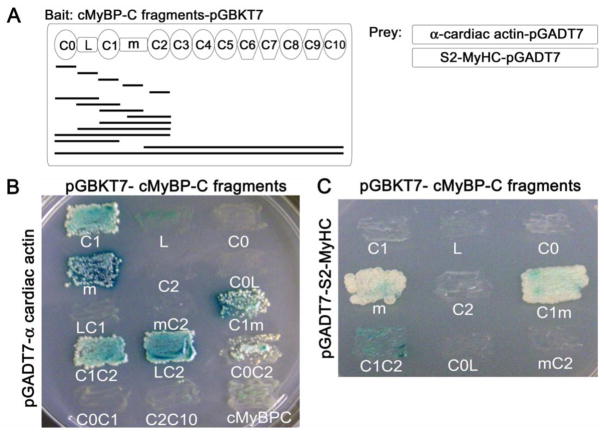
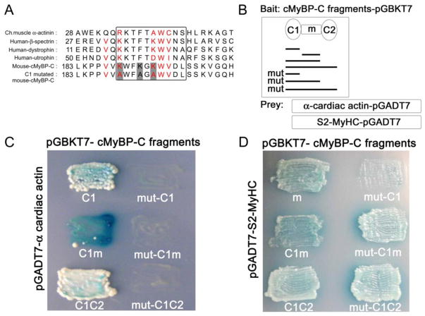
Despite early demonstrations of myosin binding protein C's (MyBP-C) interaction with actin, different investigators have reached different conclusions regarding the relevant and necessary domains mediating this binding. Establishing the detailed structure-function relationships is needed to fully understand cMyBP-C's ability to impact on myofilament contraction as mutations in different domains are causative for familial hypertrophic cardiomyopathy. We defined cMyBP-C's N-terminal structural domains that are necessary or sufficient to mediate interactions with actin and/or the head region of the myosin heavy chain (S2-MyHC). Using a combination of genetics and functional assays, we defined the actin binding site(s) present in cMyBP-C. We confirmed that cMyBP-C's C1 and m domains productively interact with actin, while S2-MyHC interactions are restricted to the m domain. Using residue-specific mutagenesis, we identified the critical actin binding residues and distinguished them from the residues that were critical for S2-MyHC binding. To validate the structural and functional significance of these residues, we silenced the endogenous cMyBP-C in neonatal rat cardiomyocytes (NRC) using cMyBP-C siRNA, and replaced the endogenous cMyBP-C with normal or actin binding-ablated cMyBP-C. Replacement with actin binding-ablated cMyBP-C showed that the mutated protein did not incorporate into the sarcomere normally. Residues responsible for actin and S2-MyHC binding are partially present in overlapping domains but are unique. Expression of an actin binding-deficient cMyBP-C resulted in abnormal cytosolic distribution of the protein, indicating that interaction with actin is essential for the formation and/or maintenance of normal cMyBP-C sarcomeric distribution.


Conflict of interest statement

Disclosure statement

The authors declare that there are no conflicts of interest.

Figures

Fig. 1
Fig. 1
Detecting functional peptide-peptide interactions between cMyBP-C and actin or cMyBP-C and S2-MyHC. (A) Shown are the N-terminal fragments derived from cMyBP-C that were used for the Y2H assays. (B) Y2H screen using cMyBP-C fragments subcloned into pGBKT7 as bait and full-length mouse α-cardiac actin subcloned into pGADT7 as prey. (C) Y2H screen using cMyBP-C fragments as bait and the S2-myosin fragment as prey identifies the m domain as being competent and sufficient to interact with S2-MyHC.
Fig. 2
Fig. 2
Identifying essential residues for protein-protein interactions. (A) Sequence alignment identifying possible homologies underlying an actin binding site. Residues K190, K193, K195 were mutated to alanines. (B) cMyBP-C’s N-terminal fragments with the C1 domain mutation used in the Y2H assays. (C) Y2H experiments showing growth/no growth on restrictive media. (D) ablation of the actin binding domain did not interfere with cMyBP-C/S2-MyHC binding.
Fig. 3
Fig. 3
Critical residues in the m domain for actin or S2-MyHC binding. (A) The m domain mutant constructs used for the Y2H assays. The residues in red were chosen for mutagenesis. (B) Y2H experiments showing mut-4 (R279A and R280A), mut-5 (R279A, R280A and T281A) and mut-8 (R304A and R305A) are not competent to mediate actin binding. (C) Mut-1 (R266A, R270A and R271A), mut-5 (R279A, R280A and T281A), mut-6 (K298A, K299A and R300A) and mut-7 (K298A, K299A) are not competent to mediate interaction with S2-MyHC. (D, E) cMyBP-C interactions with F-actin and S2-MyHC are mutually exclusive. (D) Cells were grown on SD/-His/-Leu/-Trp/X-α-gal plates: growth indicates interaction of the m domain with actin or S2-MyHC. (E) Cells plated on SD/-His/-Leu/-Met/-Trp/X-α-gal plates cannot grow, indicating that the third protein (either actin or S2-MyHC, depending upon the construct) significantly inhibited interaction between the m domain and S2-MyHC, or actin, respectively.
Fig. 4
Fig. 4
siRNA-mediated knock down of endogenous cMyBP-C. (A) Western blot analysis shows that siRNA targeted to cMyBP-C effectively decreases endogenous cMyBP-C levels. n=4, *P<0.05 (B) Immunofluorescent staining shows silencing of endogenous cMyBP-C (green). Cardiomyocytes are counterstained with a TnI antibody (red).
Fig. 5
Fig. 5
Mutant cMyBP-C expression in cardiomyocytes. (A) C1-mutated (K190A, K193A and K195A) cMyBP-C (mut-cMyBP-C) proteins in NRCs. Western blot analysis showing endogenous cMyBP-C knockdown using cMyBP-C siRNA and replaced with either normal (wt-cMyBP-C) or the mutated cMyBP-C (mut-cMyBP-C). Constructs contained a myc-tag for quantitation. Representative protein blots are shown illustrating the decreased levels of endogenous cMyBP-C and replacement to normal levels using adenovirus-driven expression of the normal or mutated cMyBP-C. Quantitation of total cMyBP-C levels (top panel, right) shows restoration of normal levels. (n=4/treatment). (B) Immunofluorescent staining with NRCs counterstained with a TnI antibody (red).

References

    1. Oakley CE, Chamoun J, Brown LJ, Hambly BD. Myosin binding protein-C: enigmatic regulator of cardiac contraction. Int J Biochem Cell Biol. 2007;39:2161–6. - PubMed
    1. Harris SP, Lyons RG, Bezold KL. In the thick of it: HCM-causing mutations in myosin binding proteins of the thick filament. Circ Res. 2011;108:751–64. - PMC - PubMed
    1. Moos C, Mason CM, Besterman JM, Feng IN, Dubin JH. The binding of skeletal muscle C-protein to F-actin, and its relation to the interaction of actin with myosin subfragment-1. J Mol Biol. 1978;124:571–86. - PubMed
    1. Kulikovskaya I, McClellan G, Flavigny J, Carrier L, Winegrad S. Effect of MyBP-C binding to actin on contractility in heart muscle. J Gen Physiol. 2003;122:761–74. - PMC - PubMed
    1. Orlova A, Galkin VE, Jeffries CM, Egelman EH, Trewhella J. The N-terminal domains of Myosin binding protein C can bind polymorphically to f-actin. J Mol Biol. 2011;412:379–86. - PMC - PubMed

Publication types