. 2012 Nov 9;423(5):766-81.
doi: 10.1016/j.jmb.2012.08.027. Epub 2012 Sep 11.

Structural analysis of the quaking homodimerization interface

Affiliations

Structural analysis of the quaking homodimerization interface

Christine Beuck et al. J Mol Biol. .

Abstract

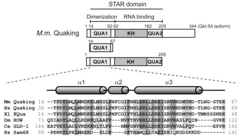
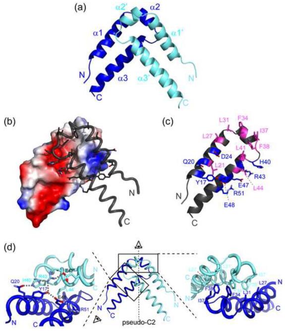
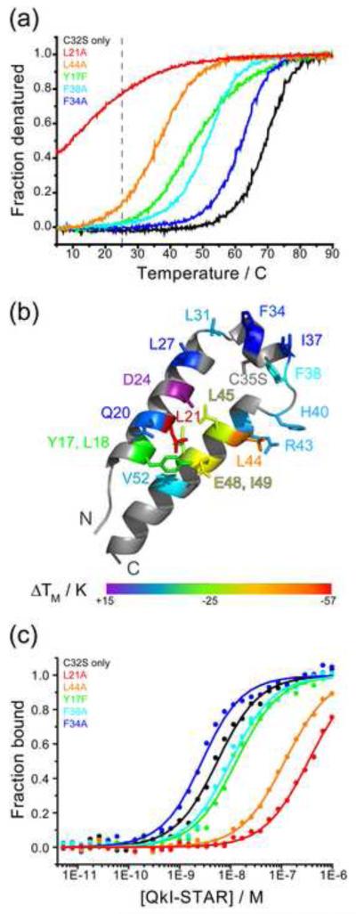
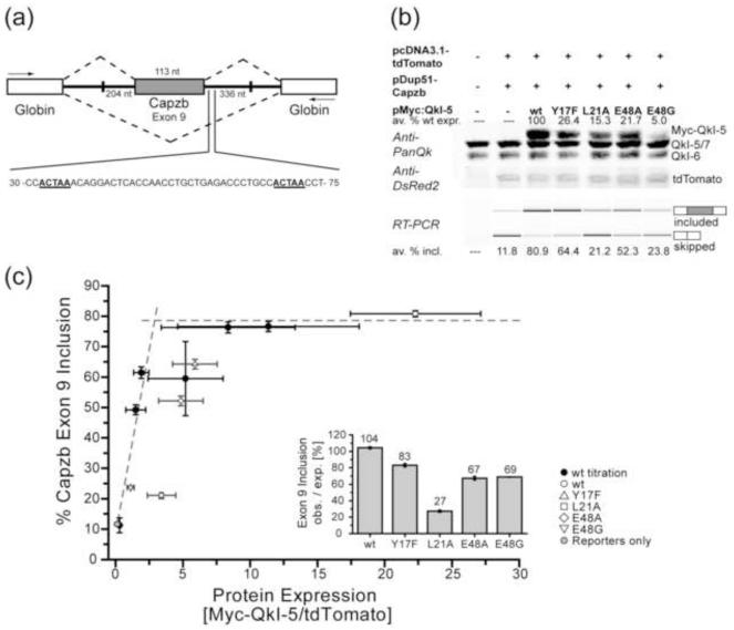
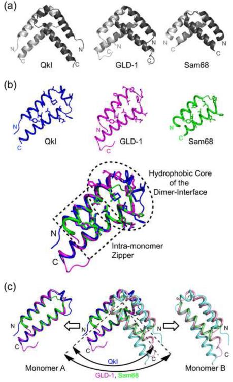
Quaking (QkI) is a prototypical member of the STAR (signal transducer and activator of RNA) protein family, which plays key roles in posttranscriptional gene regulation by controlling mRNA translation, stability and splicing. QkI-5 has been shown to regulate mRNA expression in the central nervous system, but little is known about its roles in other tissues. STAR proteins function as dimers and bind to bipartite RNA sequences; however, the structural and functional roles of homodimerization and heterodimerization are still unclear. Here, we present the crystal structure of the QkI dimerization domain, which adopts a similar stacked helix-turn-helix arrangement as its homologs GLD-1 (germ line development defective-1) and Sam68 (Src-associated protein during mitosis, 68kDa) but differs by an additional helix inserted in the dimer interface. Variability of the dimer interface residues likely ensures selective homodimerization by preventing association with non-cognate STAR family proteins in the cell. Mutations that inhibit dimerization also significantly impair RNA binding in vitro, alter QkI-5 protein levels and impair QkI function in a splicing assay in vivo. Together, our results indicate that a functional Qua1 homodimerization domain is required for QkI-5 function in mammalian cells.


Figures

Figure 1
Figure 1. The STAR family of RNA-binding proteins
Domain structure of QkI and constructs used in this study. The Qua1, KH and Qua2 subdomains are shaded (top). Sequence alignment of the Qua1 domain of representative members of the STAR/GSG protein family (bottom). Identical conserved residues are highlighted in dark grey, similar residues in light grey. The secondary structure of the Quaking Qua1 domain is shown above. Members of the Quaking subfamily feature a 3-residue insertion between the two large helices which form an additional short helix, instead of a simple turn, that is not present in the Qua1 structures of GLD-1 and Sam68.
Figure 2
Figure 2. Overall structure of the QkI-Qua1 homodimerization subdomain
(a) Structure of the QkI-Qua1 homodimer. Monomers A and B are colored in dark and light blue, respectively. (b) Dimer interface. Monomer A is shown as electrostatic surface potential, and monomer B as tube with dimer interface residues as sticks. (c) Residues of the QkI-Qua1 homodimer interface. The residues that form the hydrophobic core of the interface, containing the conserved Phe 34 and Phe 38, are highlighted in pink. Residues participating in hydrogen bonds at the edge of the interface are shown in blue. (d) Close-up view on the dimer interface. The prime denotes residues in the other protomer. Side chains of key residues in the dimer interface are shown as sticks and hydrogen bonds are indicated by grey dashed lines.
Figure 3
Figure 3. Qua1 point mutations that destabilize homodimerization also impair RNA binding
All mutants are in the C35S background and are therefore compared to QkI-Qua1 C35S. (a) Representative CD melting curves for QkI-Qua1 point mutants. Room temperature, at which the RNA binding experiments (Figure 3c) were performed, is indicated by the gray dashed line. (b) ΔTM mapped onto the monomer structure w. residues probes shown as sticks. (c) Representative RNA binding curves for QkI-STAR constructs with point mutations in the Qua1 domain. The RNA binding experiments were performed at room temperature (indicated by a grey dashed line in Figure 3a), thus only those constructs that are significantly destabilized at this temperature are expected to show a significant impact on the RNA binding affinity. The data was fit to the quadratic binding equation for bimolecular binding. The Hill equation does not apply because the RNA concentration cannot be considered ‘in trace’ (probe concentration << 10·KD) for most QkI STAR constructs.
Figure 4
Figure 4. Dimerization-deficient point mutations in the Qua1 domain reduce QkI-5 splicing activity in vivo
(a) Schematic presentation of the Capzb Exon9 mini gene organization. White boxes represent the Globin exons, the grey box represents the Capzb exon 9. Numbers by the introns flanking the Capzb Exon 9 indicate the length of intron sequence that was cloned from the Capzb gene. PCR primer positions are indicated as arrows. The 30-75 nt region downstream of the Capzb Exon 9 contains two QkI consensus binding sites, highlighted in bold and underscored, which are essential for QkI-5 splicing activity in C2C12 myoblast cells. (b) Western Blot (top) and RT-PCR analysis of QkI-5 expression levels and minigene splicing patterns from cytoplasmic RNA of C2C12 myoblast cells co-transfected with the Capzb Exon 9 minigene construct (pDup51-Capzb), plasmid for expression of wild type (wt) or mutant QkI-5 protein, and a tdTomato expression vector as transfection control. Each QkI-5 construct was assayed in three independent co-transfection experiments. In the Anti-PanQk blot, the two bottom bands represent endogenous QkI, while the top band represents the overexpressed Myc-QkI-5. The Anti-DSRed blot serves as transfection control. Average values for QkI-5 construct expression relative to wt expression are given above the Western blot. The average percentage of Capzb Exon 9 inclusion for each construct is given below the RT-PCR gel. Plots of these values including standard deviation can be found in Figure S5. (c) Capzb Exon 9 inclusion plotted versus protein expression levels. In order to normalize the splicing efficiencies of the QkI-5 mutants, which show very different protein expression levels, varying amounts of wt Myc-QkI-5 expression vector were transfected in three independent titration experiments. The protein expression level (normalized to tdTomato transfection control) and Capzb minigene splicing efficiency were analyzed as described for the QkI-5 mutants and plotted against each other. Plots of these values including standard deviation can be found in Figure S5. All individual data points were used to fit a simple saturation model (Figure S5). This plot shows the wt titration (black circles) and mutant (open symbols) data averaged over the three replicates. Error bars represent 1/2 standard deviation. The fitted saturation curve is presented as two intersecting dashed lines. Points falling below this curve indicate a lower splicing efficiency per QkI-5 molecule compared to wt, while the area above the curve indicates a higher splicing efficiency. The inset shows the ratio of observed to expected splicing efficiency (PSI, percent splicing included).
Figure 5
Figure 5. Comparison of the QkI-Qua1 structure (pdb 4DNN) to the GLD-1 (pdb 3K6T) and Sam68 (pdb 2XA6)
(a) Ribbon presentation. QkI-Qua1 features an additional 3 residues in the turn region that enable to form an additional short helix and expand the surface area of the dimer interface. (b) While the two main helices in each monomer overlay precisely for all three Qua1 domains, the two QkI-Qua1 monomers are stacked at a narrower angle compared to the GLD-1 and Sam68 homodimers. (c) Comparison of dimer interface and zipper residues (shown as sticks) of QkI, GLD-1 and Sam68 Qua1 monomers and overlay of the three structures. While the monomer zipper is highly conserved, more variation is found in the hydrophobic dimer interface residues. All three proteins use stacking of conserved Phe residues as the core of the dimer interface, but the exact position of these residues varies to some degree. The additional helix in the QkI structure broadens the dimer interface significantly and allows for more hydrogen bond interactions across the edge of the interface (see also Figure 2).

References

    1. Artzt K, Wu JI. STAR trek: An introduction to STAR family proteins and review of quaking (QKI) Adv Exp Med Biol. 2010;693:1–24. - PubMed
    1. Vernet C, Artzt K. STAR, a gene family involved in signal transduction and activation of RNA. Trends Genet. 1997;13:479–84. - PubMed
    1. Lobbardi R, Lambert G, Zhao J, Geisler R, Kim HR, Rosa FM. Fine-tuning of Hh signaling by the RNA-binding protein Quaking to control muscle development. Development. 2011;138:1783–94. - PubMed
    1. Noveroske JK, Lai L, Gaussin V, Northrop JL, Nakamura H, Hirschi KK, Justice MJ. Quaking is essential for blood vessel development. Genesis. 2002;32:218–30. - PubMed
    1. Wu JI, Reed RB, Grabowski PJ, Artzt K. Function of quaking in myelination: regulation of alternative splicing. Proc Natl Acad Sci U S A. 2002;99:4233–8. - PMC - PubMed

Publication types

Substances

LinkOut - more resources