. 2013 Jan 1:64:671-84.
doi: 10.1016/j.neuroimage.2012.09.004. Epub 2012 Sep 14.

Development of brain structural connectivity between ages 12 and 30: a 4-Tesla diffusion imaging study in 439 adolescents and adults

Affiliations

Development of brain structural connectivity between ages 12 and 30: a 4-Tesla diffusion imaging study in 439 adolescents and adults

Emily L Dennis et al. Neuroimage. .

Abstract

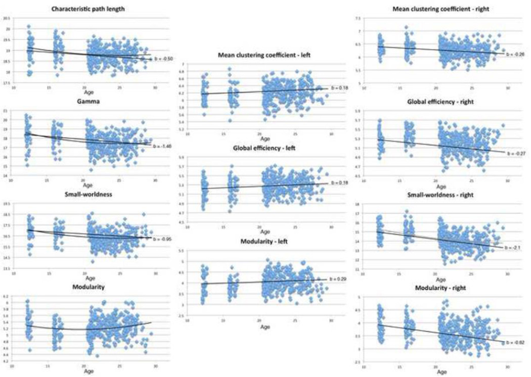
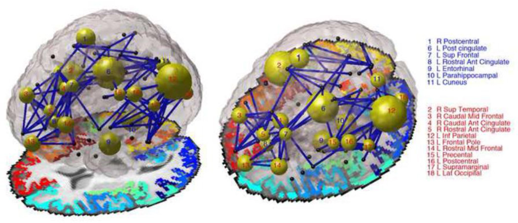
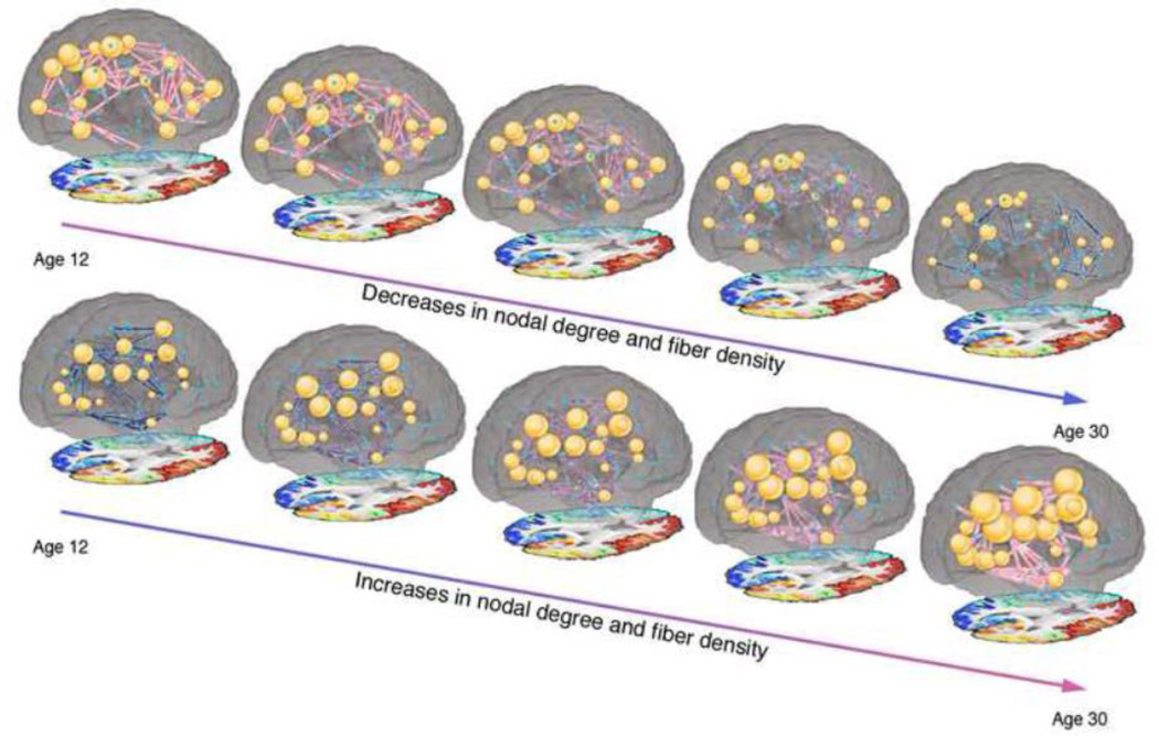
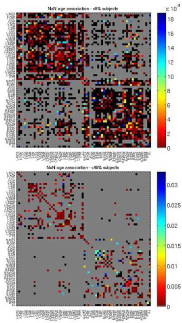
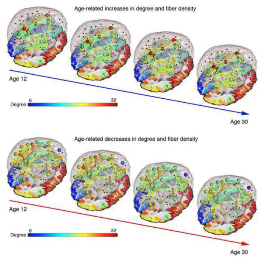
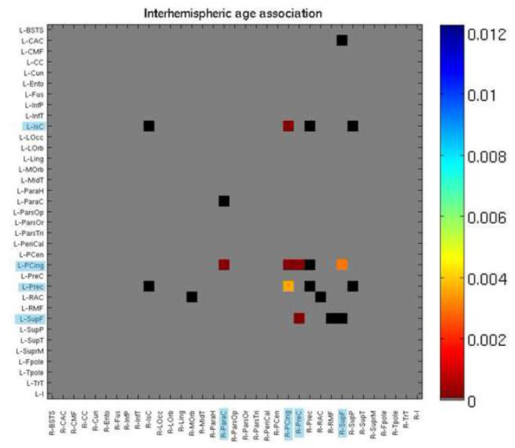
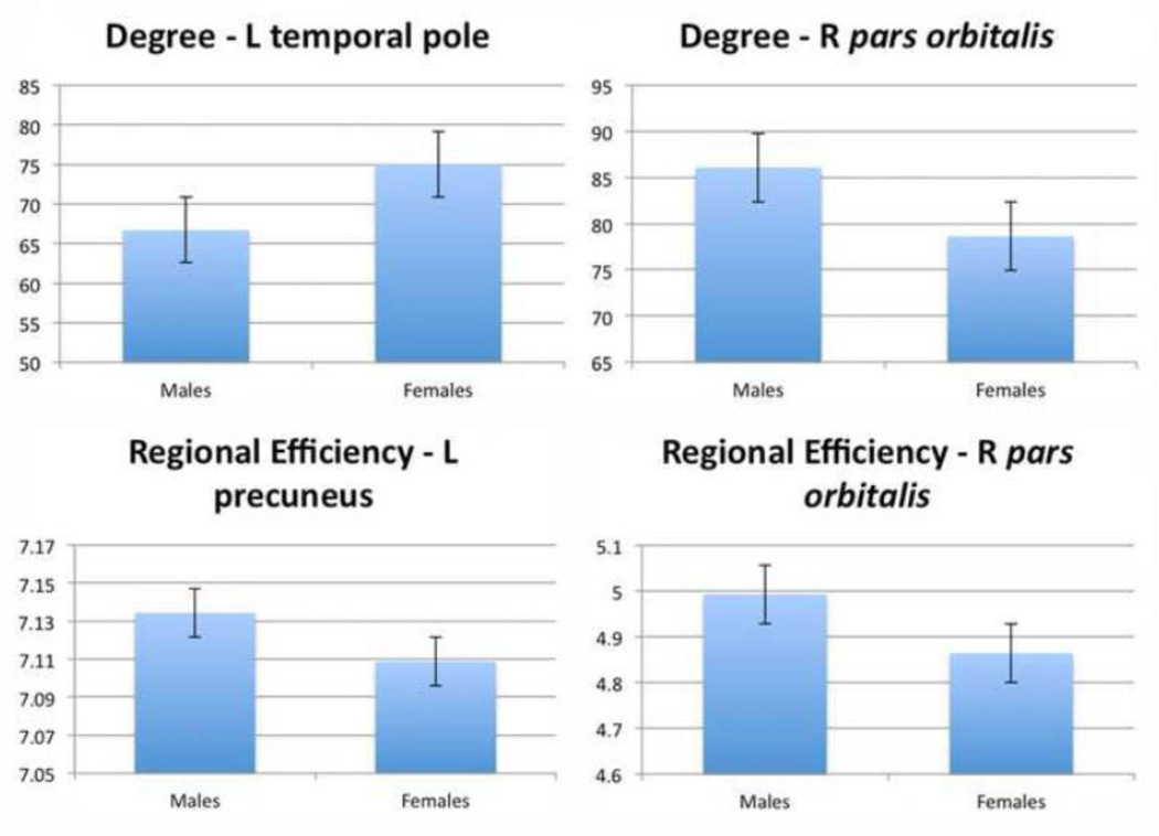
Understanding how the brain matures in healthy individuals is critical for evaluating deviations from normal development in psychiatric and neurodevelopmental disorders. The brain's anatomical networks are profoundly re-modeled between childhood and adulthood, and diffusion tractography offers unprecedented power to reconstruct these networks and neural pathways in vivo. Here we tracked changes in structural connectivity and network efficiency in 439 right-handed individuals aged 12 to 30 (211 female/126 male adults, mean age=23.6, SD=2.19; 31 female/24 male 12 year olds, mean age=12.3, SD=0.18; and 25 female/22 male 16 year olds, mean age=16.2, SD=0.37). All participants were scanned with high angular resolution diffusion imaging (HARDI) at 4 T. After we performed whole brain tractography, 70 cortical gyral-based regions of interest were extracted from each participant's co-registered anatomical scans. The proportion of fiber connections between all pairs of cortical regions, or nodes, was found to create symmetric fiber density matrices, reflecting the structural brain network. From those 70 × 70 matrices we computed graph theory metrics characterizing structural connectivity. Several key global and nodal metrics changed across development, showing increased network integration, with some connections pruned and others strengthened. The increases and decreases in fiber density, however, were not distributed proportionally across the brain. The frontal cortex had a disproportionate number of decreases in fiber density while the temporal cortex had a disproportionate number of increases in fiber density. This large-scale analysis of the developing structural connectome offers a foundation to develop statistical criteria for aberrant brain connectivity as the human brain matures.


Figures

Figure 1
Figure 1. Scatterplots showing significant associations between global graph theory connectivity scores and age in whole brain, left, and right hemispheres
Linear trendlines added with slopes and b values (regression coefficients) corresponding to results from Tables 1–3. Slopes taken from b values from Eq. 2 results, no linear trendline in included for modularity (whole brain), as that analysis was not significant.
Figure 2
Figure 2. Image depicting developmental effects, comparing children (12 and 16 year olds) to adults (20–30 year olds)
The diameter of each node is inversely proportional to the p-value for the degree analyses – large diameter means node was significantly different in degree between children and adults. Non-significant nodes are colored black. Nodes numbered in blue increase in degree with age, while those numbered with red decrease in degree with age. Blue connections are those that changed with age, corresponding to significant boxes in Figure 3. For this image we looked only at connections present in at least 95% of subjects. Author NJ is the creator of this image.
Figure 3
Figure 3
Still images from Supplementary Video 1 and Supplementary Video 2 displaying the increases and decreases in degree and fiber density between age 12 and age 30. While we lack scan data for some parts of this age range, we used the regression coefficients from our analysis to estimate network metrics at each year.
Figure 4
Figure 4. P map of age effects, when modeled alone (Eq. 2), with 70×70 fiber density matrix from which graph theory metrics were calculated
Colors correspond to strength of p value as indicated by color bar. Gray boxes were not tested as those connections were not present. For the top p map connections that were present in at least 5% of subjects were tested, for the bottom p map, connections that were present in at least 95% of subjects were tested. Black boxes were tested but not significant. FDR corrected (q < 0.05). See Table 6 for region key.
Figure 5
Figure 5. Image depicting developmental trajectory, with averaged networks shown for four groups (12 year olds, 16 year olds, 20–24 year olds, 24–30 year olds)
The color of each connection is proportional to the average fiber density within group with red signifying the thickest connections and blue the thinnest connections; the color of the node is proportional to the average degree of that node within group. For this image we looked only at connections present in at least 95% of subjects. Author NJ is the creator of this image.
Figure 6
Figure 6. P maps of age effects, when modeled alone (Eq. 2), with 35×35 interhemispheric fiber density matrix
Colors correspond to strength of p value as indicated by color bar. Blue highlighting on regions indicate significance. Gray boxes were not tested as those connections were not present. Black boxes were tested but not significant. FDR corrected (q < 0.05). See Table 6 for region key.
Figure 7
Figure 7. Bar graphs of nodes showing significant sex effects for degree (integrated over range of sparsities)
FDR corrected (q < 0.05).

References

    1. Achard S, Bullmore E. Efficiency and cost of economical brain functional networks. PLoS Comput Biol. 2007;3:e17. - PMC - PubMed
    1. Aganj I, Lenglet C, Sapiro G. Reconstruction of the orientation distribution function in single- and multiple- shell q- ball imaging within constant solid angle. Magnetic Resonance in Medicine. 2010;64(2):554–566. - PMC - PubMed
    1. Aganj I, Lenglet C, Jahanshad N, Yacoub E, Harel N, Thompson PM, Sapiro G. A Hough transform global probabilistic approach to multiple-subject diffusion MRI tractography. Medical Image Analysis. 2011;15(4):414–425. - PMC - PubMed
    1. Annett M. A classification of hand preference by association analysis. British Journal of Psychology. 1970;61(3):303–321. - PubMed
    1. Bartzokis G. Quadratic trajectories of brain myelin content: unifying construct for neuropsychiatric disorders. Neurobiology of Aging. 2004;25:49–62.

Publication types