Review
. 2012 Aug;33(3):267-86.
doi: 10.1016/j.yfrne.2012.08.006. Epub 2012 Sep 9.

The immune system and developmental programming of brain and behavior

Affiliations
Review

The immune system and developmental programming of brain and behavior

Staci D Bilbo et al. Front Neuroendocrinol. 2012 Aug.

Abstract

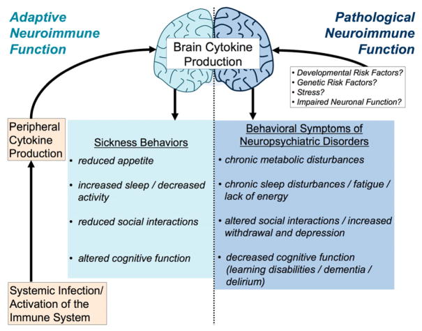
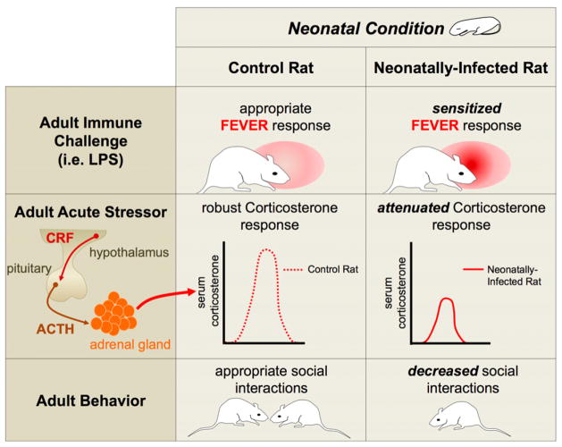
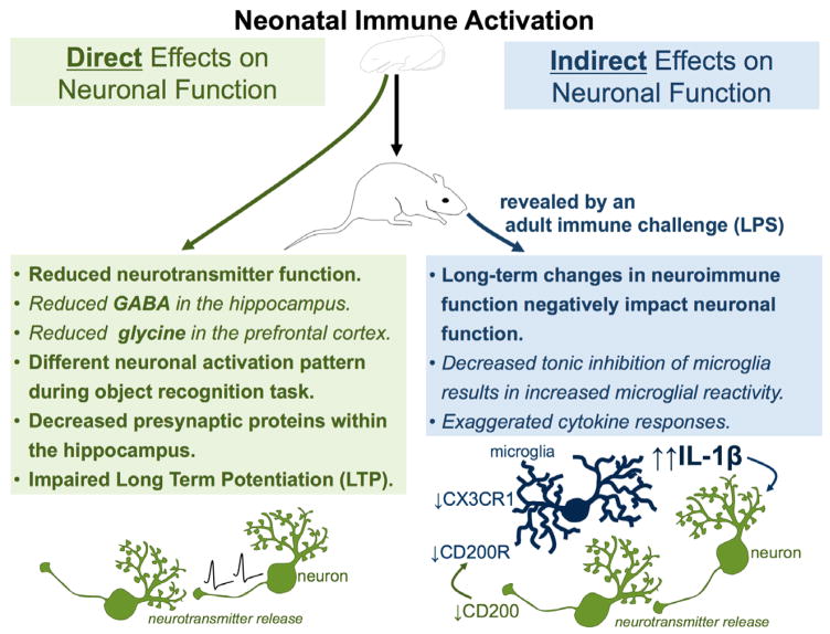
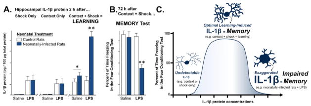
The brain, endocrine, and immune systems are inextricably linked. Immune molecules have a powerful impact on neuroendocrine function, including hormone-behavior interactions, during health as well as sickness. Similarly, alterations in hormones, such as during stress, can powerfully impact immune function or reactivity. These functional shifts are evolved, adaptive responses that organize changes in behavior and mobilize immune resources, but can also lead to pathology or exacerbate disease if prolonged or exaggerated. The developing brain in particular is exquisitely sensitive to both endogenous and exogenous signals, and increasing evidence suggests the immune system has a critical role in brain development and associated behavioral outcomes for the life of the individual. Indeed, there are associations between many neuropsychiatric disorders and immune dysfunction, with a distinct etiology in neurodevelopment. The goal of this review is to describe the important role of the immune system during brain development, and to discuss some of the many ways in which immune activation during early brain development can affect the later-life outcomes of neural function, immune function, mood and cognition.


Figures

Figure 1
Figure 1. Adaptive and pathological neuroimmune function similarly increases brain cytokine production and influences behavior
Systemic infection produces a peripheral cytokine response, which in turn produces a cytokine response in the brain. Cytokines within the brain induce a well-characterized set of adaptive behaviors that are intended to help fight infection, including reduced appetite (food and water intake), increased sleep and decreased overall activity, reduced social interactions, and altered cognitive function. Many neuropsychiatric or mood disorders exhibit a similar set of behavioral symptoms that have become prolonged or exaggerated, including chronic metabolic disorders or decreased appetite, chronic sleep disturbances/fatigue, altered social interactions, withdrawal/depression, and decreased cognitive function (e.g. learning disabilities, dementia, and delirium). Not surprisingly, many neuropsychiatric disorders are also associated with altered immune/neuroimmune function.
Figure 2
Figure 2. Neonatal infection in male rats produces a number of long-term physiological and behavioral changes
Neonatally-infected rats exhibit a sensitized fever response following an adult immune challenge, such as LPS, when compared to control rats [41]. Neonatally-infected rats have an attenuated corticosterone response to an acute stressor, when compared to control rats [42]. Neonatally-infected rats also exhibit decreased social interactions with other rats when compared to control rats [42].
Figure 3
Figure 3. Neonatal immune activation can have direct long-term effects on neuronal function or indirect long-term effects on neuronal function via alterations in neuroimmune function
Neonatal immune activation directly affects neuronal function by reducing neurotransmitter function (including GABA in the hippocampus and glycine in the prefrontal cortex), decreasing the expression of presynaptic proteins in the hippocampus, inhibiting long-term potentiation, and producing a differential neuronal activation pattern during a learning task such as the novel object recognition task. Neonatal immune activation indirectly alters neuronal function by producing long-term changes in neuroimmune function that in turn negatively impact neuronal function. Decreased tonic inhibition of microglia via altered expression of neuronal inhibitory signals, including fractalkine (via its receptor CX3CR1) and CD200, also results in exaggerated cytokine responses, which impact neuronal function.
Figure 4
Figure 4. Learning increases IL-1β protein within hippocampal microglia, which is modulated by neonatal infection
(A) Neonatally-infected rats and controls were treated in adulthood with saline or lipopolysaccharide (LPS) 24 h prior to either a learning experience (fear conditioning, consisting of 2 min context exploration followed by a footshock), or a control procedure which consisted of footshock only (without context exploration) or context exposure only (no footshock). IL-1β protein was measured in the hippocampus of separate groups of rats from each neonatal condition, 2 h after each of these conditions (shock alone, context alone, or context + shock/fear learning). In rats from both neonatal conditions that received a saline injection as adults, IL-1β protein was only increased after the learning experience (context + shock); *p<0.001, compared to context alone or shock alone. Neonatally-infected rats that received LPS 24 hours prior to behavioral testing exhibited an exaggerated IL-1β response, but only in response to learning (context + shock); **p<0.01 compared to control rats. These data indicate that normal learning induces the synthesis of IL-1β in the hippocampus, but neonatally-infected rats that receive an adult immune challenge have dysregulation of IL-1β at the time of learning. Subsequent experiments revealed that microglia were the sole source of IL-1β in these experiments [307]. (B) In a separate set of rats, fear memory for the context was assessed 72 h after conditioning. Rats from each neonatal condition that received saline 24 h prior to the learning experience show robust freezing behavior (fear) at the 72 h test, indicating that they remember the association between the shock and the context. Control rats that received LPS 24 h prior to conditioning also show robust freezing behavior (fear) at the 72 h test, indicating strong memory. In contrast, neonatally-infected rats that received LPS 24 h prior to conditioning exhibit significantly decreased freezing (fear) in the context (**p<0.05), indicating impaired memory only in this “2-hit” group (neonatal infection + adult immune challenge; see [307]). (C) Our working model is that IL-1β is produced by microglia within the hippocampus at the time of learning and is required for normal memory formation. In the absence of learning (shock alone or context alone), IL-1β is not produced in detectable levels. In neonatally-infected rats, long-term changes in neuroimmune function (microglial priming, see [307]) results in significantly exaggerated levels of IL-1β following a learning experience, which are “unmasked” by the adult LPS challenge. These exaggerated levels of IL-1β interfere with the consolidation of the learning experience and result in memory deficits. The cartoon illustrates microglia in a quiescent phenotype (left-hand tail of the inverted U), in a normal, active phenotype in which IL-1β is produced during normal learning to support memory (center), and in a sensitized/primed morphology in which exaggerated levels of IL-1β are observed in response to learning, but only in neonatally-infected rats that receive LPS as adults (right-hand tail of the inverted U). All data are represented from [307]).
Figure 5
Figure 5. Immune molecules play a ubiquitous role in neural development
Microglia, astrocytes, and neurons share a common molecular language within the CNS, and continually communicate via cytokines, chemokines, neurotransmitters and other factors (center circle). Many of the same molecules originally identified for their roles in immune function have now been implicated in neural development; representative examples are conceptualized here (see [48, 81] for comprehensive reviews). (1) Many cytokines are important for progenitor cell maintenance, proliferation, and differentiation. The bone-morphogenic protein (BMP)/transforming growth factor beta (TGFβ) family of cytokines is critical for neural induction [106]. The gp130 receptor and associated ligands are important for progenitor cell maintenance and proliferation [80, 117, 175]. TLR3 is important for maintaining progenitor cell populations and proliferation of dividing cells within the brain [157], and HMGB1, a known ligand for TLR4, impacts cell survival [319]. (2) Cytokines such as IL-1β and the IL-6 family of proteins have a demonstrated role in cytogenesis within the developing brain [50, 198]. IL-1β expression peaks during astrocytogenesis, which is dependent on the presence of amoeboid microglia [109]. Microglia begin to colonize the developing brain as primitive yolk sac macrophages beginning around E9–10 [107]; macrophage colony-stimulating factor (M-CSF) and the CSF-1 receptor are important for this recruitment [107, 250]. (3) Chemokines, in particular CXCL12 and its receptor CXCR4, guide the migration of new neurons in many brain regions, including the cerebellum [166, 321]. (4) MHC I is critical for the activity dependent formation of synapses within the visual cortex, and likely many other brain regions [60, 78, 261]. TNFα released by astrocytes promotes synaptic transmission and affects activity-dependent synaptic scaling [270]. (5) Complement proteins, C1q and C3, tag synapses for elimination [272]. Microglia recognize these proteins via the complement receptor 3 (CD11b), and phagocytose the labeled synapses as a mechanism of synaptic pruning [254]. (6) Microglia are the primary phagocytic cells of the CNS, and thus have an important role in phagocytosing apoptotic debris following programmed cell death, a process that occurs continuously and most abundantly within the developing brain [93]. Programmed cell death likely plays a major role in recruitment of microglia into the CNS [302].
Figure 6
Figure 6. The list of factors hypothesized to activate immune cells via Toll-Like Receptors (TLRs), in particular TLR4
Toll-like receptors are innate pattern recognition receptors which identify the pathogen-associated molecular patterns (PAMPs) of specific pathogens. For example, TLR4 recognizes the immunologically active cell wall pattern of gram-negative bacteria, lipopolysaccharide (LPS), which is also found on E. coli. The role of these receptors has recently expanded to include the broader recognition of “danger” associated molecular patterns (DAMPs). These include a novel subset of proteins that are produced and released by nearby cells undergoing cell death or distress, known as alarmins, which are putatively recognized by TLRs, including both TLR4 and TLR2. Proteins that have been identified as alarmins thus far include hyaluronan, Heat Shock Proteins, and high mobility group box (HMGB) 1. In addition, the list of exogenous or environmental factors that are hypothesized to activate TLR4 (directly or indirectly) is ever-expanding. These include drugs of abuse (e.g. opiates, amphetamines, and ethanol), saturated dietary fats, and other toxins (e.g. air pollution).

References

    1. Adams-Chapman I, Stoll BJ. Neonatal infection and long-term neurodevelopmental outcome in the preterm infant. Current opinion in infectious diseases. 2006;19:290–297. - PubMed
    1. Aiuti A, Webb IJ, Bleul C, Springer T, Gutierrez-Ramos JC. The chemokine SDF-1 is a chemoattractant for human CD34+ hematopoietic progenitor cells and provides a new mechanism to explain the mobilization of CD34+ progenitors to peripheral blood. The Journal of experimental medicine. 1997;185:111–120. - PMC - PubMed
    1. Alexopoulou L, Holt AC, Medzhitov R, Flavell RA. Recognition of double-stranded RNA and activation of NF-kappaB by Toll-like receptor 3. Nature. 2001;413:732–738. - PubMed
    1. Anderson KV, Jurgens G, Nusslein-Volhard C. Establishment of dorsal-ventral polarity in the Drosophila embryo: genetic studies on the role of the Toll gene product. Cell. 1985;42:779–789. - PubMed
    1. Andersson U, Tracey KJ. Reflex principles of immunological homeostasis. Annual Review of Immunology. 2012;30:313–335. - PMC - PubMed

Publication types

MeSH terms