. 2013 Feb:65:134-42.
doi: 10.1016/j.neuropharm.2012.09.004. Epub 2012 Sep 13.

Thalamic glutamatergic afferents into the rat basolateral amygdala exhibit increased presynaptic glutamate function following withdrawal from chronic intermittent ethanol

Affiliations

Thalamic glutamatergic afferents into the rat basolateral amygdala exhibit increased presynaptic glutamate function following withdrawal from chronic intermittent ethanol

Daniel T Christian et al. Neuropharmacology. 2013 Feb.

Abstract

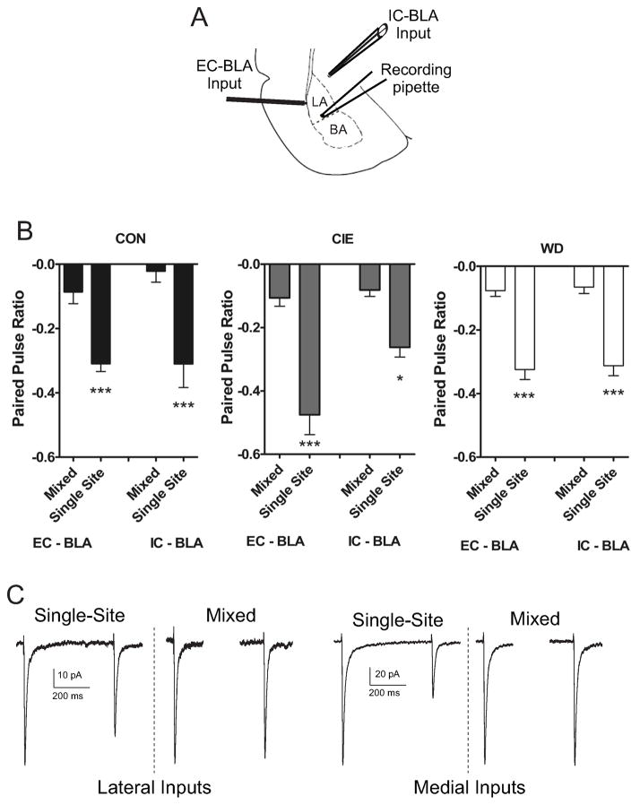
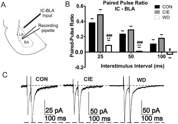
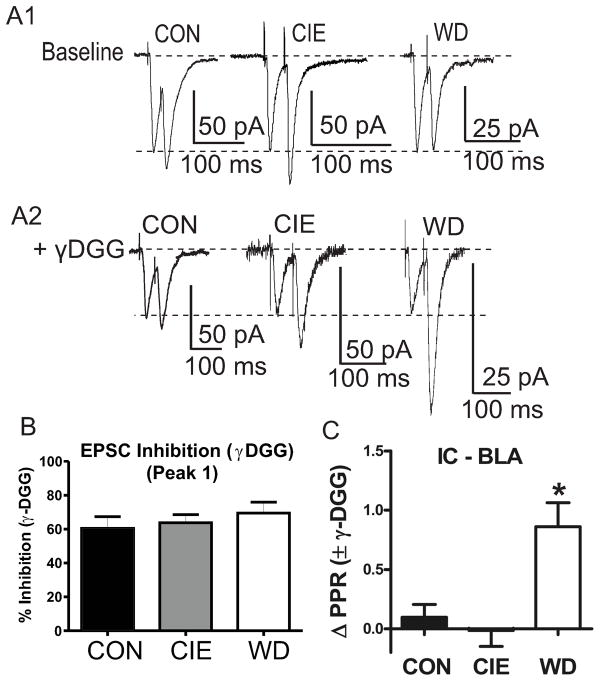
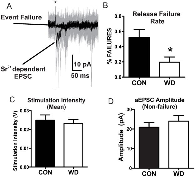
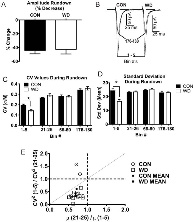
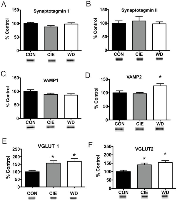
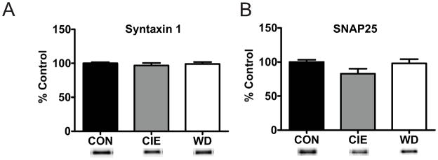
Amygdala glutamatergic neurotransmission regulates withdrawal induced anxiety-like behaviors following chronic ethanol exposure. The lateral/basolateral amygdala receives multiple glutamatergic projections that contribute to overall amygdala function. Our lab has previously shown that rat cortical (external capsule) afferents express postsynaptic alterations during chronic intermittent ethanol exposure and withdrawal. However, thalamic (internal capsule) afferents also provide crucial glutamatergic input during behavioral conditioning, and they have not been studied in the context of chronic drug exposure. We report here that these thalamic inputs express altered presynaptic function during withdrawal from chronic ethanol exposure. This is characterized by enhanced release probability, as exemplified by altered paired-pulse ratios and decreased failure rates of unitary events, and increased concentrations of synaptic glutamate. Quantal analysis further implicates a withdrawal-dependent enhancement of the readily releasable pool of vesicles as a probable mechanism. These functional alterations are accompanied by increased expression of vesicle associated protein markers. These data demonstrate that chronic ethanol modulation of glutamate neurotransmission in the rat lateral/basolateral amygdala is afferent-specific. Further, presynaptic regulation of lateral/basolateral amygdala thalamic inputs by chronic ethanol may be a novel neurobiological mechanism contributing to the increased anxiety-like behaviors that characterize withdrawal.


Figures

Figure 1
Figure 1. Anatomically distinct glutamatergic BLA afferents are functionally distinct across CIE and WD
A) Diagram representing relative placement of recording and stimulating electrode placements in External Capsule (EC) and Internal Capsule (IC) BLA afferents for electrophysiology experiments (adopted from (Paxinos and Watson, 1997)). B) Paired electrical stimuli (500 ms) were delivered to EC or IC afferents in either a single afferent (EC-EC; IC-IC) or mixed afferent (EC-IC; IC-EC). Paired, single site stimulation elicited robust, but similar paired pulse depression at both afferents across all treatment conditions that did not [(EC1 – EC2), Kruskal-Wallis = 5.34, p > 0.05; (IC1 – IC2) Kruskal-Wallis = 0.36, p > 0.05]. Mixed afferent stimulation elicited little paired pulse depression that did not differ across treatment [(EC1– EC2), Kruskal-Wallis = 1.13, p > 0.05; (IC1 – IC2), Kruskal-Wallis = 2.43, p > 0.05]. ANOVA (treatment condition vs. stimulation site) indicated a significant main effect for stimulation site [EC- EC paired, (−0.37 ± 0.02 n = 18); EC - EC unpaired, (−0.08 ± 0.01 n = 18); IC - IC paired, (−0.29 ± 0.02 n = 18); IC - IC unpaired (−0.05 ± 0.01 n = 18); F(3,60) = 48.748, p < 0.05] but not treatment [CON (−0.18 ± 0.03, n = 24); CIE (−0.23 ± 0.03, n = 24); WD (−0.19 ± 0.02, n = 24); F(2,60) = 1.84, p > 0.05] with no significant interaction [F (6,60) = 1.781, p > 0.05]. This lack of functional interaction indicates that these anatomically distinct afferent pathways are functionally distinct, independent of treatment. C) Representative traces showing paired pulse depression during single afferent stimulation at both EC/IC afferents. Mixed afferent stimulation results in little paired pulse depression at either input.
Figure 2
Figure 2. WD increases presynaptic function at IC-BLA afferents
A) Diagram representing relative placements of stimulating (IC-BLA) and recording electrodes (BLA) for electrophysiology experiments. B) Decreased PPRs during WD indicate increased release probability at multiple interstimulus intervals: 25ms [CON, 0.37 ± 0.05, n = 16; CIE, 0.48 ± 0.06, n = 10; WD, 0.08 ± 0.06, n = 11; F(2,34) = 9.464, p < 0.05], 50ms [CON, 0.23 ± 0.04, n = 16; CIE, 0.28 ± 0.04, n = 14; WD, 0.01 ± 0.05, n = 15; F(2,42) = 9.488 p > 0.05], 100ms [CON, 0.10 ± 0.05, n = 15; CIE, 0.17 ± 0.04, n = 13, WD, −0.02 ± 0.04 n =15; F(2,40) = 4.117 p > 0.05)], One Way ANOVA’s, Newman-Keuls post hoc tests. ### = significant vs. CON; # ,** = significant vs. CIE. C) Representative traces of PPR recordings (50 ms) scaled to second peak amplitude during each treatment condition.
Figure 3
Figure 3. WD increases synaptic glutamate concentration
A1) Representative traces of baseline paired pulse stimulation (25 ms) across treatment conditions. Traces are scaled to first response amplitudes in all cases. A2) Representative traces following bath application of γDGG (1.0 mM) illustrate decreased second pulse inhibition by γDGG during WD. B) Inhibition of first-response amplitude by γDGG was not different across treatment groups [CON 60.49 ± 6.81%, n =7; CIE, 63.70 ± 4.82, n = 10; WD, 69.48 ± 6.49, n =7; F(2,21) = .052; p > 0.05]. C) WD slices exhibit significantly greater PPR values following bath application of γDGG due to decreased pulse to pulse inhibition [ΔPPR; CON 0.09 ± 0.10, n = 7; CIE, −0.01 ± 0.13, n = 10; WD, 0.86 ± 0.20, n = 7; F(2,21) = 9.475, p < 0.05 Newman Keuls Post Hoc Test * = significant from CON and CIE].
Figure 4
Figure 4. Decreased rates of vesicle release failures during WD
A) Representative traces showing mean Sr2+ dependent EPSC responses and non release event failures (Black traces). Representative raw data (20 sweeps) from exemplar cell (Gray traces). * indicates truncated stimulus artifact. B) WD decreases release failure rates [CON (8); WD (7); t = 2.492, df = 13, * = p < 0.05]. C) Stimulation intensities did not differ between treatment conditions [CON (8); WD (7); t = 0.47 df = 13, p > 0.05]. D) Non-failure EPSC amplitude events do not differ with treatment [CON (8), WD (7); t =0.824, df = 13, p > 0.05].
Figure 5
Figure 5. CV analysis localizes WD dependent functional alterations to presynaptic compartments
A) Absolute EPSC rundown are not different between groups (t = 0.17, df = 14, p > 0.05). B) Representative traces scaled to Bin 1–5 peak values show no change in mean amplitudes. C) CV values (σ/M) decrease in WD (n=8) relative to CON (n=8) during initial rundown [Bin 1–5; t = 4.30 df = 8, * = p < 0.05], but not across extended time points [bins 21–25, t = 1.741, df = 8; bins 56–60, t = 0.02, df = 8; bins 176–180, t = 0.70, df = 8; p > 0.05 for all comparisons]. D) WD decreases σ values during Bins 1–5 [bins 1–5, t = 3.88, df = 8, * = p < 0.05] but not at extended Bin intervals [bins 21–25, t = 0.21, df = 8, p > 0.05; bins 56–60, t = 1.06, df = 8, p > 0.05; bins 176–180, t = 0.03, df = 8, p > 0.05]. E) CV2 analysis characterizing localization of increased WD dependent glutamate function with values on or below the diagonal line suggesting a presynaptic mechanism of action.
Figure 6
Figure 6. WD dependent alterations of vesicle proteins during WD
A, B) Protein expression of Synaptotagmin I [CON, 100.00 ± 4.72, n = 8; CIE, 87.47 ± 4.56, n = 9; WD, 98.11 ± 4.51, n = 8] and II [CON, 100.00 ± 8.83, n = 4; CIE, 108.38 ± 17.33; n = 5; WD, 97.73 ± 7.33, n = 4] are unaltered following WD. C) VAMP1 protein expression is unaltered by CIE or WD [100.00 ± 6.16, n = 12; CIE, 88.99 ± 4.17, n = 12; WD, 86.44 ± 4.61, n = 12]. D) WD but not CIE increases VAMP2 protein expression [CONE, 100.00 ± 7.33, n = 11; CIE, 96.70 ± 4.48, n = 12; WD, 125.68 ± 7.96, n = 12]. E, F) VGlut protein expression increases during both CIE and WD VGlut1: [CON, 100.00 ± 10.48, n = 8; CIE, 157.56 ± 15.31, n = 9; WD, 168.98 ± 17.83, n = 9; F(2,23) = 5.697, p < 0.05 * indicates significant difference from CON]; VGlut2: [CON, 100.00 ± 8.74, n = 8; CIE, 140.24 ± 10.80, n = 9; WD, 154.32 ± 10.82, n = 9; F(2,23) = 7.226, p < 0.05 * indicates significant difference from CON)]. Increased protein expression of vesicle associated protein suggest in increase in vesicle size or number. VAMP2 and VGlut2 protein data suggests a pathway specific increase in synaptic vesicles.
Figure 7
Figure 7. T-Snare protein is not regulated by CIE or WD
A) Total protein expression of T-Snare Syntaxin 1 is unaffected by CIE and WD exposures [CON, 100.00 ± 1.64, n =8; CIE, 96.75 ± 3.71, n = 8; WD, 99.12 ± 2.79, n = 8; F (2,21) = 0.348; p > 0.05]. B) T-Snare SNAP25 protein expression is unchanged following CIE and WD exposures [CON, 100.00 ± 3.26, n = 8; CIE, 82.86 ± 7.33, n = 8; WD, 97.94 ± 6.24, n = 8; F (2,21) = 2.537, p > 0.05]. No treatment dependent alterations in T-Snare protein expression levels suggest that the number of release sites at presynaptic terminals is unchanged.

Similar articles

Cited by

References

    1. Bauer EP, Schafe GE, LeDoux JE. NMDA receptors and L-type voltage-gated calcium channels contribute to long-term potentiation and different components of fear memory formation in the lateral amygdala. J Neurosci. 2002;22:5239–5249. - PMC - PubMed
    1. Becherer U, Rettig J. Vesicle pools, docking, priming, and release. Cell and Tissue Research. 2006;326:393–407. - PubMed
    1. Bender VA, Pugh JR, Jahr CE. Presynaptically Expressed Long-Term Potentiation Increases Multivesicular Release at Parallel Fiber Synapses. The Journal of Neuroscience. 2009;29:10974–10978. - PMC - PubMed
    1. Boatman JA, Kim JJ. A thalamo-cortico-amygdala pathway mediates auditory fear conditioning in the intact brain. European Journal of Neuroscience. 2006;24:894–900. - PubMed
    1. Budzinski KL, Allen RW, Fujimoto BS, Kensel-Hammes P, Belnap DM, Bajjalieh SM, Chiu DT. Large Structural Change in Isolated Synaptic Vesicles upon Loading with Neurotransmitter. Biophysical Journal. 2009;97:2577–2584. - PMC - PubMed

Publication types

MeSH terms

LinkOut - more resources