

Utility of GATA3 immunohistochemistry in differentiating urothelial carcinoma from prostate adenocarcinoma and squamous cell carcinomas of the uterine cervix, anus, and lung
- PMID: 22982890
- PMCID: PMC3444740
- DOI: 10.1097/PAS.0b013e318260cde7
Utility of GATA3 immunohistochemistry in differentiating urothelial carcinoma from prostate adenocarcinoma and squamous cell carcinomas of the uterine cervix, anus, and lung
Abstract
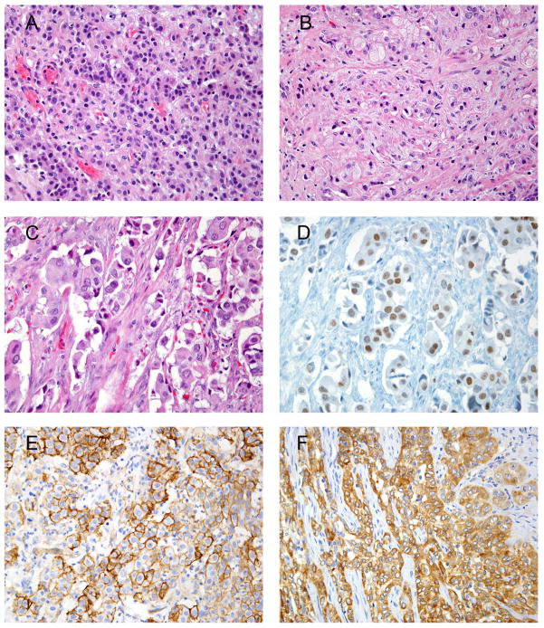
Distinguishing invasive high-grade urothelial carcinoma (UC) from other carcinomas occurring in the genitourinary tract may be difficult. The differential diagnosis includes high-grade prostatic adenocarcinoma, spread from an anal squamous cell carcinoma (SCC), or spread from a uterine cervical SCC. In terms of metastatic UC, the most common problem is differentiating spread of UC to the lung from a primary pulmonary SCC. Immunohistochemical analysis (IHC) for GATA binding protein 3 (GATA3), thrombomodulin (THROMBO), and uroplakin III was performed on a tissue microarray (TMA) containing 35 cases of invasive high-grade UC. GATA3 IHC was also performed on TMAs containing 38 high-grade (Gleason score ≥8) prostatic adenocarcinomas, representative tissue sections from 15 invasive anal SCCs, representative tissue sections from 19 invasive cervical SCCs, and TMAs with 12 invasive cervical carcinomas of the cervix [SCC (n=10), SCC with neuroendocrine features (n=1), and adenosquamous carcinoma (n=1)]. In addition, GATA3 IHC was performed on representative tissue sections from 15 pulmonary UC metastases and a TMA with 25 SCCs of the lung and 5 pulmonary non-small cell carcinomas with squamous features. GATA3, THROMBO, and uroplakin III were positive in 28 (80%), 22 (63%), and 21 (60%) cases of high-grade UC, respectively. All cases of GATA3-positive staining were nonfocal; 25 (89%) cases demonstrated moderate to strong staining, and 3 (11%) demonstrated weak staining. Of the 7 cases that failed to express GATA3, 5 were positive for THROMBO and/or uroplakin III, whereas 2 were negative for all 3 markers. None of the 38 high-grade prostatic adenocarcinomas was positive for GATA3. Weak GATA3 staining was present in occasional basal cells of benign prostate glands, in a few benign atrophic glands, and in urothelial metaplasia. Of the 15 cases of anal SCCs, 2 (7%) cases showed focal weak staining, and 1 (3%) showed focal moderate staining. Weak staining was also rarely observed in the benign anal squamous epithelium. Of the 31 uterine cervical carcinomas, 6 (19%) showed weak GATA3 staining (3 nonfocal and 3 focal), and 2 (6%) demonstrated focal moderate staining. Twelve (80%) of the metastatic UCs to the lung were positive for GATA3, with 11 cases showing diffuse moderate or strong staining and 1 case showing focal moderate staining. None of the pulmonary SCCs or non-small cell carcinomas with squamous features was GATA3 positive. GATA3 IHC is a sensitive marker for UC, and positive staining in UC is typically nonfocal and moderate or strong in intensity. GATA3 is also highly specific in excluding high-grade prostate adenocarcinoma. Although some cervical and anal SCCs can be GATA3 positive, unlike in UC, staining is more commonly focal and weak. GATA3 is also a useful maker when diagnosing metastatic UC to the lung.
Conflict of interest statement
Disclosures: The authors have no conflicts of interest or relevant funding to disclose.
Figures
References
-
- Asselin-Labat ML, Sutherland KD, Barker H, et al. Gata-3 is an essential regulator of mammary-gland morphogenesis and luminal-cell differentiation. Nat Cell Biol. 2007;9:201–209. - PubMed
-
- Chuang AY, DeMarzo AM, Veltri RW, et al. Immunohistochemical differentiation of high-grade prostate carcinoma from urothelial carcinoma. Am J Surg Pathol. 2007;31:1246–1255. - PubMed
-
- Esheba GE, Longacre TA, Atkins KA, et al. Expression of the urothelial differentiation markers GATA3 and placental S100 (S100P) in female genital tract transitional cell proliferations. Am J Surg Pathol. 2009;33:347–353. - PubMed
-
- Gaisa NT, Braunschweig T, Reimer N, et al. Different immunohistochemical and ultrastructural phenotypes of squamous differentiation in bladder cancer. Virchows Arch. 2011;458:301–312. - PubMed
MeSH terms
Substances
Grants and funding
LinkOut - more resources
Full Text Sources
Medical
Research Materials
