. 2012 Aug;223(1):7-20.
doi: 10.1086/BBLv223n1p7.

Investigating bacterial-animal symbioses with light sheet microscopy

Affiliations

Investigating bacterial-animal symbioses with light sheet microscopy

Michael J Taormina et al. Biol Bull. 2012 Aug.

Abstract

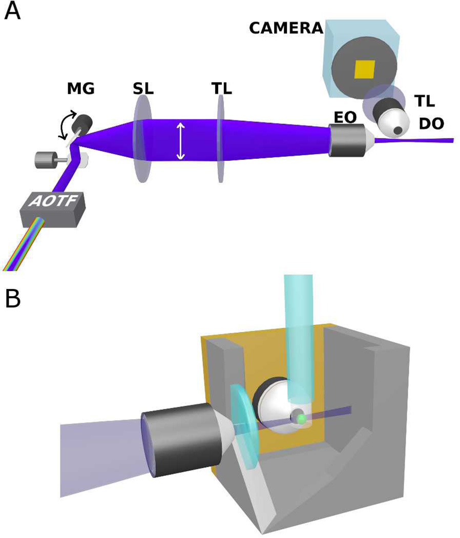
Microbial colonization of the digestive tract is a crucial event in vertebrate development, required for maturation of host immunity and establishment of normal digestive physiology. Advances in genomic, proteomic, and metabolomic technologies are providing a more detailed picture of the constituents of the intestinal habitat, but these approaches lack the spatial and temporal resolution needed to characterize the assembly and dynamics of microbial communities in this complex environment. We report the use of light sheet microscopy to provide high-resolution imaging of bacterial colonization of the intestine of Danio rerio, the zebrafish. The method allows us to characterize bacterial population dynamics across the entire organ and the behaviors of individual bacterial and host cells throughout the colonization process. The large four-dimensional data sets generated by these imaging approaches require new strategies for image analysis. When integrated with other "omics" data sets, information about the spatial and temporal dynamics of microbial cells within the vertebrate intestine will provide new mechanistic insights into how microbial communities assemble and function within hosts.


Figures

Figure 1
Figure 1
A 7 dpf zebrafish larva. A brightfield image (A) and a schematic representation (B) illustrating the intestinal tract. sb indicates the swim bladder and grey fill highlights the intestine. Scale bar: 0.5 mm.
Figure 2
Figure 2
Schematic illustrations of fluorescence imaging techniques: (A) wide-field, (B) point scanning confocal, (C) multi-photon, and (D) light sheet microscopy. In all diagrams, points A and B lie in the focal plane of the lens while point C lies outside the focal plane. Optical sectioning in light sheet microscopy works by creating a plane of excitation light that is coincident with the focal plane of an imaging objective. Since the sectioning occurs during excitation, the emitted light from the whole field of view can be gathered as a two-dimensional image with a standard camera.
Figure 3
Figure 3
(A) Schematic illustration of our light sheet microscope setup. Abbreviations: AOTF, acousto-optic tunable filter; MG, mirror galvanometer; SL, scan lens; TL, tube lens; EO, excitation objective lens; DO, detection objective lens. (B) Schematic illustration of our specimen holder, in which the EO axis, DO axis, and capillary tube are all mutually perpendicular. The green sphere denotes the specimen, which is embedded in agarose gel and held by a glass capillary from above.
Figure 4
Figure 4
Colonization of a larval zebrafish gut by GFP-expressing and dTomato-expressing Aeromonas veronii bacteria. (A) wild type GFP-expressing and dTomato-expressing A. veronii coinoculated in culture. Panels B–J show representative 2D optical sections from 3D data sets. (B) The entire gut six hours after inoculation. The gut is approximately outlined with a dashed line and the swim bladder (sb) is outlined with a solid line. (C–J) The distribution of the two microbial populations in the region corresponding to the box in (B) at various times after inoculation, with t = 6 corresponding to panel B. The contrast of each panel was independently adjusted so that the bacterial populations are clearly visible despite fluctuations in overall bacterial abundance. In each panel, both red and green intensities were rescaled by the same amount. (J) The intensity rescaling factor in each of panels (B–I), i.e. the sum of the red and green intensities each normalized by the typical brightness of a typical bacterium, providing a measure of the total bacterial abundance at each time. Scale bars: (A) 10 microns (B–J) 100 microns
Figure 5
Figure 5
(A) Fluorescently labeled enteroendocrine cells (transgenic line nkx2.2a:egfp) in a larval zebrafish gut. Scale bar: 40 microns. (B–G) A time series of the cells within the box in (A). The interval between each panel in (B–G) is 2.8 seconds. The deformation and movement of cells as the gut undergoes peristalsis during the imaging period is evident. The movie from which these images were taken is provided as Supplemental Movie S2. (H–M) A single fluorescently labeled neutrophil (mpo:gfp) within the intestinal tissue of a larval zebrafish. Each panel is a two-dimensional optical section from a three-dimensional data set, each separated in time by 2 minutes. Dynamic rearrangments of the neutrophil’s filopodia are evident. Scale bar: 10 microns.
Figure 6
Figure 6
Population distributions as a function of time for the two co-inoculated bacterial populations illustrated in Figure 4. Each column indicates fluorescence intensity integrated over the dimensions perpendicular to the gut axis, providing a one-dimensional measure of bacterial density along the gut, the temporal dynamics of which can be visualized in a two-dimensional plot.
Figure 7
Figure 7
Image auto- and cross-correlations. (A) A simulated two-dimensional image with randomly positioned green spots and 1.5× larger red spots each placed approximately 20 pixels from a green spot, randomly oriented. (B) The two-dimensional cross-correlation, Cgr, of the green and red channels of (A); a ring at a radius of 20 pixels reveals the constructed correlation. (C) The radial dependence of the auto- and cross-correlation functions; as in (B), Cgr shows a peak at a spatial offset of 20 pixels. (D) A simulated two-dimensional image in which both the green and red spots are positioned randomly. (E, F) the correlation functions corresponding to the image in (D). (G) A single optical slice from a three-dimensional data set depicting Aeromonas veronii bacteria in a larval zebrafish, as in Figure 4, at a single time point. Scale bar: 50 microns. (H) Auto- and cross-correlations of the bacterial intensity distributions calculated from the three-dimensional data set.

References

    1. Altarriba M, Merino S, Gavin R, Canals R, Rabaan A, Shaw JG, Tomas JM. A polar flagella operon (flg) of Aeromonas hydrophila contains genes required for lateral flagella expression. Microb Pathog. 2003;34:249–259. - PubMed
    1. Bates JM, Akerlund J, Mittge E, Guillemin K. Intestinal alkaline phosphatase detoxifies lipopolysaccharide and prevents inflammation in zebrafish in response to the gut microbiota. Cell Host Microbe. 2007;2:371–382. - PMC - PubMed
    1. Bates JM, Mittge E, Kuhlman J, Baden KN, Cheesman SE, Guillemin K. Distinct signals from the microbiota promote different aspects of zebrafish gut differentiation. Dev Biol. 2006;297:374–386. - PubMed
    1. Bjornstad ON, Ims RA, Lambin X. Spatial population dynamics: analyzing patterns and processes of population synchrony. Trends Ecol Evol. 1999;14:427–432. - PubMed
    1. Boettcher KJ, Ruby EG, McFall-Ngai MJ. Bioluminescence in the symbiotic squid Euprymna scolopes is controlled by a daily biological rhythm. Journal of Comparative Physiology A Sensory Neural and Behavioral Physiology. 1996;179:65–73.

Publication types

LinkOut - more resources