. 2012 Nov 1;303(9):L748-57.
doi: 10.1152/ajplung.00102.2012. Epub 2012 Sep 14.

TLR4 deficiency promotes autophagy during cigarette smoke-induced pulmonary emphysema

Affiliations

TLR4 deficiency promotes autophagy during cigarette smoke-induced pulmonary emphysema

Chang Hyeok An et al. Am J Physiol Lung Cell Mol Physiol. .

Abstract

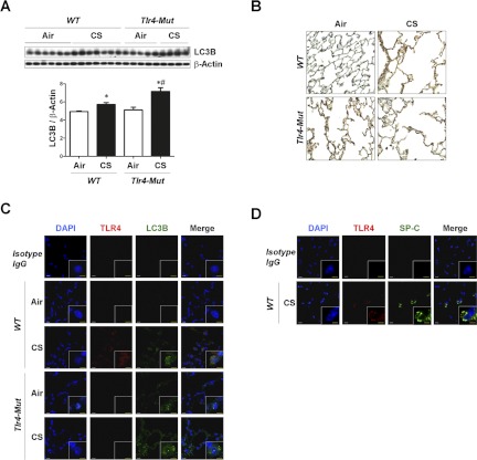
Toll-like receptors (TLRs) exert important nonimmune functions in lung homeostasis. TLR4 deficiency promotes pulmonary emphysema. We examined the role of TLR4 in regulating cigarette smoke (CS)-induced autophagy, apoptosis, and emphysema. Lung tissue was obtained from chronic obstructive lung disease (COPD) patients. C3H/HeJ (Tlr4-mutated) mice and C57BL/10ScNJ (Tlr4-deficient) mice and their respective control strains were exposed to chronic CS or air. Human or mouse epithelial cells (wild-type, Tlr4-knockdown, and Tlr4-deficient) were exposed to CS-extract (CSE). Samples were analyzed for TLR4 expression, and for autophagic or apoptotic proteins by Western blot analysis or confocal imaging. Chronic obstructive lung disease lung tissues and human pulmonary epithelial cells exposed to CSE displayed increased TLR4 expression, and increased autophagic [microtubule-associated protein-1 light-chain-3B (LC3B)] and apoptotic (cleaved caspase-3) markers. Beas-2B cells transfected with TLR4 siRNA displayed increased expression of LC3B relative to control cells, basally and after exposure to CSE. The basal and CSE-inducible expression of LC3B and cleaved caspase-3 were elevated in pulmonary alveolar type II cells from Tlr4-deficient mice. Wild-type mice subjected to chronic CS-exposure displayed airspace enlargement;, however, the Tlr4-mutated or Tlr4-deficient mice exhibited a marked increase in airspace relative to wild-type mice after CS-exposure. The Tlr4-mutated or Tlr4-deficient mice showed higher levels of LC3B under basal conditions and after CS exposure. The expression of cleaved caspase-3 was markedly increased in Tlr4-deficient mice exposed to CS. We describe a protective regulatory function of TLR4 against emphysematous changes of the lung in response to CS.


Figures

Fig. 1.
Fig. 1.
Toll-like receptors 4 (TLR4) is induced in patients with an advanced stage of chronic obstructive lung disease (COPD) in parallel with increases in autophagic protein light-chain-3B (LC3B) and apoptosis. Human lung tissues of never-smokers and patients with COPD Global Initiative for Obstructive Lung Disease (GOLD) stage 2 (G2) and 4 (G4) were analyzed by Western blot analysis (n = 5 for never-smokers; n = 15 for patients with COPD GOLD stage 2; n = 15 for patients with COPD GOLD stage 4). Western blot analysis for TLR4 and cleaved caspase-3 were quantified for relative TLR4 (A), and cleaved caspase-3 (B) expression. β-Actin served as the standard. Data are means ± SE; *P < 0.05 compared with never-smoker; #P < 0.05 compared with previous group. Western blot analysis of LC3B was quantified for LC3B-II (C). β-Actin served as the standard. Data are means ± SE; *P < 0.05 compared with never-smoker; #P < 0.05 compared with previous group. D: representative images of human lung tissues with immunohistochemical staining for LC3B. E: immunofluorescence staining for TLR4 (red) and LC3B (green). Scale bar indicates 10 μm. F: immunofluorescence staining for TLR4 (red) and SP-C (green) as the marker of type II alveolar epithelial cells. DAPI, 4,6-diamidino-2-phenylindole. Scale bar = 10 μm.
Fig. 2.
Fig. 2.
Increased TLR4 expression regulates autophagic protein LC3B and apoptosis in cigarette smoke-extract (CSE)-treated epithelial cells. A: Beas-2B cells were treated with CSE (20%) for the indicated times, and their cell lysates were analyzed by Western blot analysis for TLR4 and TLR2. β-Actin served as the standard. B: A549 cells were treated with CSE (20%) for the indicated times, and their cell lysates were analyzed by Western blot analysis for TLR4 and TLR2. β-Actin served as the standard. C: human bronchial epithelial (HBE) cells were treated with CSE (10%) for the indicated times and their cell lysates were analyzed by Western blot analysis for TLR4, LC3B, and cleaved caspase-3. β-Actin served as the standard. D: Beas-2B cells were treated with CSE (20%) for the indicated times and their cell lysates were analyzed by Western blot analysis for TLR4 and LC3B. β-Actin served as the standard. E: Beas-2B cells were pretreated with control siRNA (CTL) or TLR4-siRNA (TLR4) for 48 h, followed by CSE (20%) treatment for 6 h. The lysates were then subjected to Western blot analysis as indicated. β-Actin served as the standard. F: primary alveolar type II (ATII) cells were isolated from C57BL/10ScNJ (Tlr4-deficient) and C57BL/10SnJ [wild-type (WT)] mice. ATII cells were exposed to CSE (20%) for 6 h and the lysates were immunoblotted for TLR4, LC3B, and cleaved caspase-3. β-Actin served as the standard. G: primary ATII cells were isolated from C57BL/10SnJ (WT) mice and cultured in air-liquid interface (ALI) state. ATII cells were exposed to 50 mg/m3 of total particulate matter from one cigarette for 10 min and the lysates were immunoblotted for TLR4 and LC3B. β-Actin served as the standard. H: ATII cells were treated with CSE (20%) for the indicated times, and the culture media was subjected to lactate dehydrogenase (LDH) release assay. Data represent means ± SE; *P < 0.05 compared with preceding day in same mouse group; #P < 0.05 compared with wild-type mice at same day.
Fig. 3.
Fig. 3.
Pulmonary emphysema was augmented by exposure to CS in Tlr4-mutated or Tlr4-deficient mice. A: quantification of mean linear intercepts (MLIs) of C3H/HeOuJ (WT) and C3H/HeJ (Tlr4-mutated) mice exposed to CS for 2 mo. Data represent means ± SE; *P < 0.05 compared with air-exposed mice in the same strain; #P < 0.05 compared with WT mice with same exposure. B: representative images of lungs are shown with modified Gill's staining. C: quantification of MLIs of C56BL/10SnJ (WT) and C56BL/10ScNJ (Tlr4-deficient) mice exposed to CS for 6 mo. Data represent means ± SD; *P < 0.05 compared with air-exposed mice in the same strain; #P < 0.05 compared with WT mice with same exposure. D: representative images of lungs are shown with modified Gill's staining.
Fig. 4.
Fig. 4.
TLR4 regulates CS-induced autophagic protein LC3B and apoptosis in vivo. C57BL/10SnJ (WT) or C57BL/10ScNJ (Tlr4-deficient) mice were exposed to CS or air for 6 mo (C57BL/10SnJ, n = 6 for air, n = 5 for CS; C57BL/10ScNJ, n = 5 for air, n = 4 for CS). Western blot analysis and corresponding quantification for LC3B (A) and cleaved caspase-3 (B). Data are means ± SE; *P < 0.05 compared with air-exposure group in the same strain; #P < 0.05 compared with WT mice with same exposure. C: representative images of immunohistochemical staining for LC3B in mouse lung tissues. D: immunofluorescence staining for TLR4 (red) and LC3B (green) in mouse lung tissues. E: immunofluorescence staining for TLR4 (red) and SP-C (green) as a marker of ATII cells in mouse lung tissues. White scale bar = 10 μm; yellow scale bar = 5 μm. F: lipid peroxidation [malondialdehyde (MDA)] assay of homogenates of lung tissues. Data are means ± SE; *P < 0.05 compared with air-exposure group in the same strain; #P < 0.05 compared with WT mice with same exposure. G: immunofluorescence staining for 4-hydroxy-2-nonenal (4-HNE) (red) as a marker of lipid peroxidation in mouse lung tissues. Blue indicates DAPI stain for nuclear staining.
Fig. 5.
Fig. 5.
C3H/HeOuJ (WT) or C3H/HeJ (Tlr4-mutated) mice were exposed to CS or air for 2 mo (C3H/HeOuJ, n = 6 for air, n = 6 for CS; C3H/HeJ, n = 5 for air, n = 5 for CS). Western blot analysis and its corresponding quantification (A) and immunohistochemical staining (B) for LC3B were performed in mouse lungs. Data represent means ± SE; *P < 0.05 compared with air-exposure group in the same strain; #P < 0.05 compared with WT mice with same exposure. C: immunofluorescence staining for TLR4 (red) and LC3B (green) in mouse lung tissues. D: immunofluorescence staining for TLR4 (red) and SP-C (green) as a marker of ATII cells in mouse lung tissues. White scale bar = 10 μm; yellow scale bar = 5 μm.

References

    1. Barnes PJ. Chronic obstructive pulmonary disease. N Engl J Med 343: 269–280, 2000 - PubMed
    1. Bertin A, Samson M, Pons C, Guigonis JM, Gavelli A, Baqué P, Brossette N, Pagnotta S, Ricci JE, Pierrefite-Carle V. Comparative proteomics study reveals that bacterial CpG motifs induce tumor cell autophagy in vitro and in vivo. Mol Cell Proteomics 7: 2311–2322, 2008 - PubMed
    1. Bishai JM, Mitzner W. Effect of severe calorie restriction on the lung in two strains of mice. Am J Physiol Lung Cell Mol Physiol 295: L356–L362, 2008 - PMC - PubMed
    1. Chen ZH, Kim HP, Sciurba FC, Lee SJ, Feghali-Bostwick C, Stolz DB, Dhir R, Landreneau RJ, Schuchert MJ, Yousem SA, Nakahira K, Pilewski JM, Lee JS, Zhang Y, Ryter SW, Choi AM. Egr-1 regulates autophagy in cigarette smoke-induced chronic obstructive pulmonary disease. PLoS One 3: e3316, 2008 - PMC - PubMed
    1. Chen ZH, Lam HC, Jin Y, Kim HP, Cao J, Lee SJ, Ifedigbo E, Parameswaran H, Ryter SW, Choi AM. Autophagy protein microtubule-associated protein 1 light chain-3B (LC3B) activates extrinsic apoptosis during cigarette smoke-induced emphysema. Proc Natl Acad Sci USA 107: 18880–18885, 2010 - PMC - PubMed

Publication types

MeSH terms