. 2013 Mar;14(1):15-22.
doi: 10.1007/s10195-012-0213-z. Epub 2012 Sep 16.

Lumbar spine MRI in upright position for diagnosing acute and chronic low back pain: statistical analysis of morphological changes

Affiliations

Lumbar spine MRI in upright position for diagnosing acute and chronic low back pain: statistical analysis of morphological changes

Umberto Tarantino et al. J Orthop Traumatol. 2013 Mar.

Abstract

Background: Patients with low back pain frequently demonstrate recumbent magnetic resonance imaging (MRI) alterations not always related to homogeneous clinical symptoms. The purpose of this study was to evaluate and quantify the statistical significance of variations of some anatomical parameters of the lumbosacral spine and reveal occult disc pathologies from recumbent to upright position in patients with acute and chronic low back pain.

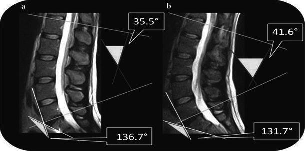
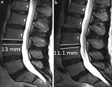
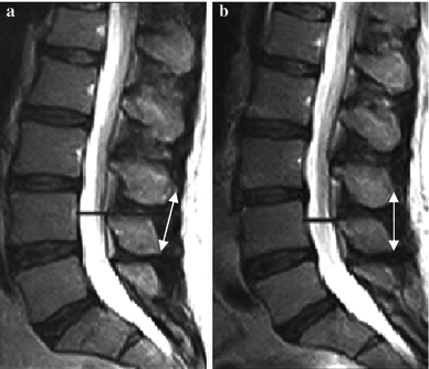
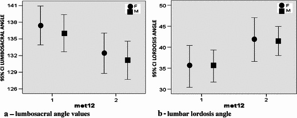
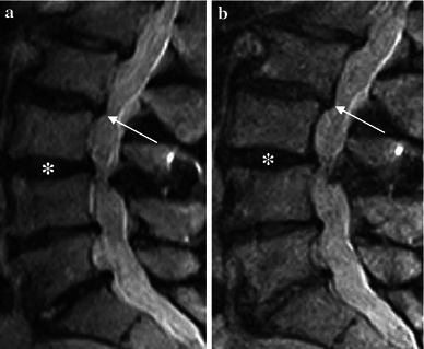
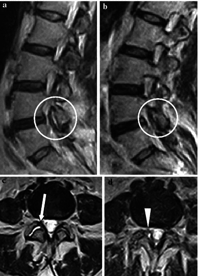
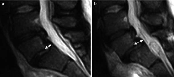
Materials and methods: Fifty-seven patients complaining of low back pain (27 women, 30 men) underwent dynamic lumbosacral MRI with a 0.25-T tilting system (G-scan Esaote). We settled five parameters for which variations have been evaluated: lumbosacral angle, lordosis angle, L3-L4 intersomatic disc height, L3-L4 interspinous processes distance, and widest anteroposterior dural sac diameter. Images were obtained in both recumbent and upright positions.

Results: Statistically significant differences [one-way analysis of variance (ANOVA), p = 0.0043] were found between each pair of values of parameters sampled in recumbent and upright positions. In 70 % of patients, on visual qualitative analysis only, an increment of disc protrusions and/or spondylolisthesis was found in the upright position; in three cases, in the upright position only, an interarticular pseudocyst was found.

Conclusions: Dynamic MRI with an open-configuration, low-field tilting MRI system is a feasible and promising tool to study degenerative pathology of the spine. Moreover, in cases of low back pain with negative MRI in the recumbent position or in patients with pain in the upright position only, tilting MRI permits visualization of occult spine and disc pathologies in patients with acute or chronic low back pain.


Figures

Fig. 1
Fig. 1
Fast spin echo (FSE) T2-weighted magnetic resonance images (MRI) in the sagittal plane. Lumbosacral angle and lumbar lordosis angle are average values. a Supine position: lumbosacral angle 136.7°, lordosis angle 35.5°. b Upright position: lumbosacral angle 131.7°, lordosis angle 41.6°
Fig. 2
Fig. 2
Fast spin echo (FSE) T2-weighted magnetic resonance images (MRI) in the sagittal plane. Assessment of changes in thickness of the intervertebral disc in the transition from a supine to b upright position, with averaged values
Fig. 3
Fig. 3
Fast spin echo (FSE) T2-weighted magnetic resonance images (MRI) in the sagittal plane. Assessment of changes in interspinous distance (white arrows) and amplitude of the dural sac (black bars) in the transition from a clinostatism to b orthostasis
Fig. 4
Fig. 4
Statistical distribution: Changes in a lumbosacral and b lordosis angle. a Clinostatism: lumbosacral 136.7°, lordosis 35.5°. b Orthostasis: lumbosacral 131.7°, lordosis 41.6°
Fig. 5
Fig. 5
Statistical distribution of the intersomatic disc thickness between sexes. Average value in clinostatism 12.9 mm and orthostasis 11.2 mm
Fig. 6
Fig. 6
Statistical analysis of variation of the interspinous distance between sexes. Average value in clinostatism 14.6 mm and orthostasis 12.8 mm
Fig. 7
Fig. 7
Statistical analysis of amplitude variation of the dural sac between sexes. Average value in clinostatism 13.1 mm and orthostasis 14.5 mm
Fig. 8
Fig. 8
Fast spin echo (FSE) T2-weighted magnetic resonance images (MRI) in the sagittal plane. Disc protrusions in the entire L1–S1 section: a Supine, b standing. Slight accentuation of the protruding component in the upright position between L2 and L3 (arrows) and reduced discal height in the interspace between L3 and L4 (asterisks) is visible
Fig. 9
Fig. 9
Fast spin echo (FSE) T2-weighted magnetic resonance images (MRI) in the sagittal and axial planes. a, c Clinostatism; b, d orthostasis. c Presence of a fluid collection between articular facets at L4–L5 (arrow). d Orthostatic position shows evagination of pseudocystic appearAnce of the right joint capsule with an impression on the nerve root and dural sac (arrowhead)
Fig. 10
Fig. 10
Fast spin echo (FSE) T2-weighted magnetic resonance images (MRI) in the sagittal plane. a Clinostatism; b orthostasis. Listhesis is accentuated in the upright position passing from grade I to II and making disc protrusion more evident

Similar articles

Cited by

References

    1. Gallucci M, Limbucci N, Paonessa A, Splendiani A. Degenerative disease of the spine. Neuroimaging Clin of N Am. 2007;17:87–103. doi: 10.1016/j.nic.2007.01.002. - DOI - PubMed
    1. Splendiani A, Di Fabio MV, Barile A, Masciocchi C. Tecnica di studio RM del rachide lombare sotto carico. In: Leone A, Martino F, editors. Imaging del rachide. Il vecchio e il nuovo. Italy: Springer; 2008. pp. 123–134.
    1. Hoy D, Brooks P, Blyth F, Buchbinder R. The epidemiology of low back pain. Best Pract Res Clin Rheumatol. 2010;24:769–781. doi: 10.1016/j.berh.2010.10.002. - DOI - PubMed
    1. Claus A, Hides J, Moseley GL, Hodges P. Sitting versus standing: does the intradiscal pressure cause disc degeneration or low back pain? J Electromyogr Kinesiol. 2008;18:550–558. doi: 10.1016/j.jelekin.2006.10.011. - DOI - PubMed
    1. Parent EC, Videman T, Battié MC. The effect of lumbar flexion and extension on disc contour abnormality measured quantitatively on magnetic resonance imaging. Spine (Phila Pa 1976) 2006;31:2836–2842. doi: 10.1097/01.brs.0000245834.30646.aa. - DOI - PubMed