. 2012 Oct 12;338(6104):226-31.
doi: 10.1126/science.1222538. Epub 2012 Sep 13.

Cilia at the node of mouse embryos sense fluid flow for left-right determination via Pkd2

Affiliations

Cilia at the node of mouse embryos sense fluid flow for left-right determination via Pkd2

Satoko Yoshiba et al. Science. .

Abstract

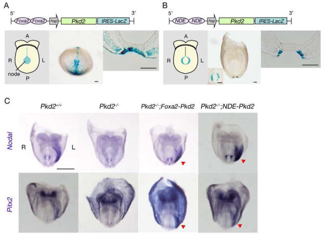
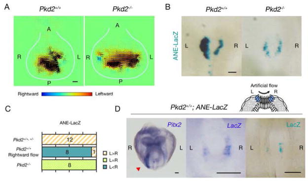
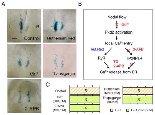
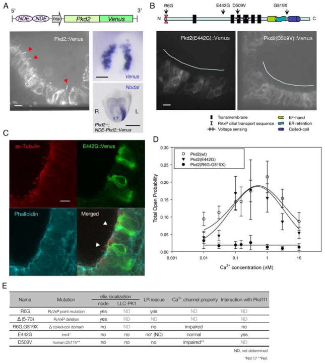
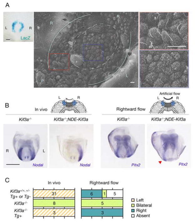
Unidirectional fluid flow plays an essential role in the breaking of left-right (L-R) symmetry in mouse embryos, but it has remained unclear how the flow is sensed by the embryo. We report that the Ca(2+) channel Polycystin-2 (Pkd2) is required specifically in the perinodal crown cells for sensing the nodal flow. Examination of mutant forms of Pkd2 shows that the ciliary localization of Pkd2 is essential for correct L-R patterning. Whereas Kif3a mutant embryos, which lack all cilia, failed to respond to an artificial flow, restoration of primary cilia in crown cells rescued the response to the flow. Our results thus suggest that nodal flow is sensed in a manner dependent on Pkd2 by the cilia of crown cells located at the edge of the node.


Figures

Figure 1
Figure 1. Specific requirement for Pkd2 in crown cells of the node for correct L-R patterning
(A, B) Schematic representation of two types of node-specific Pkd2 transgeneare shown on the top. Their expression patterns were examined by staining of transgenic embryos at embryonic day (E) 8.0 with X-gal. Schematic (left panels) and actual (middle panels) views of whole embryos, and transverse sections of the node (right panels) are shown. Inset of the middle panel in B shows node from ventral view at higher magnification. Note that expression of the NDE-driven transgene is highly specific to crown cells of the node (B). A, anterior; P, posterior; L, left; R, right. Scale bars, 50μm. (C) Whole-mount in situ hybridization analysis of the expression of Nodal and Pitx2 in E8.0 embryos of the indicated genotypes. Left-sided expression of Nodal and Pitx2 in LPM is lost in Pkd2−/− embryos but is restored (red arrowheads) in Pkd2−/−;Foxa2-Pkd2 and Pkd2−/−;NDE-Pkd2 embryos. Scale bar, 500μm.
Figure 2
Figure 2. Pkd2 is necessary for sensing of nodal flow
(A) Nodal flows in wild-type and Pkd2−/− embryos were examined by PIV analysis at E8.0. Flow is maintained in the Pkd2−/− embryo. Each arrowhead denotes the direction and speed of the flow at the indicated position. The color scale indicates the direction and magnitude of the flow velocity (leftward in yellow and red, rightward in blue). White lines indicate the outlines of the node. Scale bar, 10μm. (B) E8.0 Pkd2+/+ and Pkd2−/− embryos harboring the ANE-LacZ transgene were stained with X-gal. Scale bar, 50μm. (C) The L-R patterns of ANE activities in embryos of the indicated genotypes either in situ or under conditions of artificial rightward flow in vitro are summarized. The numbers of embryos showing each pattern are indicated. (Adjust the color/pattern for C. The small box definitions of colors will not show the pattern of top line after figure reduction. ---response: color/patterns have been changed and it is easy to see the difference in color/pattern) (D) Pkd2+/+ embryos with ANE-LacZ were cultured under the influence of a rightward artificial flow from early headfold to six-somite stages. Artificial rightward flow reversed the pattern of Pitx2 expression in LPM (left panel, red arrowhead) and that of ANE activity in crown cells, as detected by in situ hybridization of LacZ mRNA (middle) and X-gal staining (right). Scale bars, 100μm. Why does the “L” and “R” designation switch from left and right between the figure panels? Figure 1 shows “L” on the right side of figure; Figure 2 shows “L” and “R” in different panel sides in the figure. ---Response: embryos showing gene expression in the lateral plate such as ones in Fig. 1C are frontal views, so that “L ” is shown on the right side. Embryos showing gene expression in the node such as ones in Fig. 2B were back views, so that “L ” was shown on the left side. To avoid confusion, however, all embryo photos are shown in such a way that the right side of an embryo is always shown on the left side.) (Define N and C.---response: N & C are now deleted)
Figure 3
Figure 3. Calcium signal is essential for L-R symmetry breaking in the node crown cells
(A) Activity of ANE is disrupted when Ca2+ signal blockers are treated. Two pathways that lead to Ca2+ release from endoplasmic reticulum were examined (B). L>R asymmetric ANE activity is maintained in control and Ruthenium Red- treated embryos, whereas it is disrupted with Gd3+, 2-aminoethoxydiphenyl borate (2-APB) or Thapsigargin (TG). Scale bar, 50μm. (C) The effects of each reagent on L-R asymmetric ANE activity are summarized. The numbers of embryos showing each pattern are indicated.
Figure 4
Figure 4. Ciliary localization of Pkd2 is required for correct L-R determination
(A) Schematic representation of NDE-driven Pkd2::Venus transgene is shown on the top. Crown cell-specific expression of the transgene in an E8.0 embryo was confirmed by whole-mount in situ hybridization with a Venus probe (upper right panel). Scale bar, 50μm. Left-sided expression of Nodal was restored in Pkd2−/−; NDE-Pkd2::Venus embryo (lower right panel), suggesting that Pkd2::Venus is functional. Scale bar, 500μm. Note that Pkd2::Venus protein is preferentially localized to cilia (left panel, red arrowheads). Scale bar, 10 μm. (B) Transgenic embryos expressing Pkd2(E442G)::Venus or Pkd2(D509)::Venus in crown cells. Note that both proteins are unable to localize to cilia. Scale bar, 10 μm. (Define N and C. ---response: N & C are removed) (C) Localization of Pkd2(E442G)::Venus was confirmed by immunofluorescence staining with antibodies to acetylated tubulin (red—difficult to see/read tubulin---response: we have changed the color brighter so that it is easier to see), to Venus; GFP (green) and phalloidin (cyan). Scale bar, 10μm. (D) Open probability of Pkd2(wt), Pkd2(E442G) and Pkd2(R6G-G819X) channels in the presence of increasing cytosolic Ca2+ concentration. Note that Pkd2(E442G) retains normal channel activity whereas Pkd2(R6G-G819X) loses it. Error bars represent ± SEM. (E) The relationship between ciliary localization, Ca2+ channel activity, ability of L-R rescue and interaction with Pkd1l1 is summarized for various Pkd2 mutants. (Please remove E from the figures section and make it a separate Table.---response: we have made E a separate Table).
Figure 5
Figure 5. Nodal flow is sensed by the cilia of crown cells
(A) An E8.0 transgenic embryo harboring NDE-Kif3a-IRES-LacZ was stained with X-gal, revealing crown cell specific expression of the transgene (left). Scale bar, 50μm. In scanning electron microscopy of the node of an E8.0 Kif3a−/− embryo with NDE-Kif3a, cilia are apparent at the edge (boxed by the red line), but not at the center (boxed by the blue line) of the node. Pale blue lines indicate the border between the endoderm and crown cells, with the dotted circle enclosing pit cells. The boxed regions on the left are shown at a higher magnification on the right. Scale bars, 5 μm. (B) Expression of L-R marker genes in embryos of the indicated genotypes that were examined at E8.0 (in vivo) or cultured under the influence of a rightward artificial flow before analysis. Note that Pitx2 expression pattern of the Kif3a−/−;NDE-Kif3a embryo responded to the flow and is right-sided (red arrowhead). Scale bar, 500 μm. (C) The numbers of embryos showing each pattern of gene expression are summarized.

Comment in

References

    1. Shiratori H, Hamada H. Development. 2006;133:2095. - PubMed
    1. Hirokawa N, Tanaka Y, Okada Y. Cold Spring Harb Perspect Biol. 2009;1:a000802. - PMC - PubMed
    1. Gonzalez-Perrett S, et al. Proc Natl Acad Sci U S A. 2001;98:1182. - PMC - PubMed
    1. Koulen P, et al. Nat Cell Biol. 2002;4:191. - PubMed
    1. Pennekamp P, et al. Curr Biol. 2002;12:938. - PubMed

Publication types

MeSH terms