. 2013 May;36(3):499-512.
doi: 10.1007/s10545-012-9530-x. Epub 2012 Sep 15.

Retroviral-vector-mediated gene therapy to mucopolysaccharidosis I mice improves sensorimotor impairments and other behavioral deficits

Affiliations

Retroviral-vector-mediated gene therapy to mucopolysaccharidosis I mice improves sensorimotor impairments and other behavioral deficits

Guilherme Baldo et al. J Inherit Metab Dis. 2013 May.

Abstract

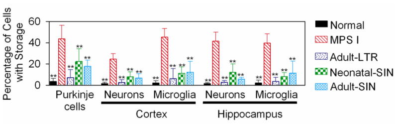
Mucopolysaccharidosis I (MPS I) is a lysosomal storage disease due to α-L-iduronidase (IDUA) deficiency that results in the accumulation of glycosaminoglycans (GAG). Systemic gene therapy to MPS I mice can reduce lysosomal storage in the brain, but few data are available regarding the effect upon behavioral function. We investigated the effect of gene therapy with a long-terminal-repeat (LTR)-intact retroviral vector or a self-inactivating (SIN) vector on behavioral function in MPS I mice. The LTR vector was injected intravenously to 6-week-old MPS I mice, and the SIN vector was given to neonatal or 6-week-old mice. Adult-LTR, neonatal-SIN, and adult-SIN-treated mice achieved serum IDUA activity of 235 ± 20 (84-fold normal), 127 ± 10, and 71 ± 7 U/ml, respectively. All groups had reduction in histochemical evidence of lysosomal storage in the brain, with the adult-LTR group showing the best response, while adult-LTR mice had reductions in lysosomal storage in the cristae of the vestibular system. Behavioral evaluation was performed at 8 months. Untreated MPS I mice had a markedly reduced ability to hold onto an inverted screen or climb down a pole. LTR-vector-treated mice had marked improvements on both of these tests, whereas neonatal-SIN mice showed improvement in the pole test. We conclude that both vectors can reduce brain disease in MPS I mice, with the LTR vector achieving higher serum IDUA levels and better correction. Vestibular abnormalities may contribute to mobility problems in patients with MPS I, and gene therapy may reduce symptoms.


Figures

Fig. 1
Fig. 1. Serum α-L-iduronidase (IDUA) activity
A. Serum IDUA activity after treatment in MPS I mice. Mice were injected with 1.7×10¹⁰ TU/kg of the RV designated hAAT-cIDUA-WPRE at 1.5 months of age (Adult-LTR), with 1×10¹⁰ TU/kg of the self-inactivating version of the RV designated SIN-hAAT-cIDUA-oPRE at 2 to 3 days after birth (Neonatal-SIN), or with 1×10¹⁰ TU/kg of the SIN vector at 1.5 months of age (Adult-SIN) and serum IDUA ± standard deviation (SD) was measured using a fluorogenic substrate as described in the methods section from 1 week until 8 months of age. Results represent the average of the lifetime average for the indicated number of mice in each group. B. Forebrain and cerebellar IDUA activity. Animals were sacrificed at 8 months and IDUA levels were measured in the forebrain and cerebellum for 5 normal, 6 untreated MPS I, 4 Neonatal-SIN, and 3 Adult-SIN mice, and from forebrain of 6 Adult-LTR mice; some treated mice were not evaluated due to lack of sample availability. The number of mice of each gender was similar. C. β-glucuronidase (GUSB) activity. The X over the position of the cerebellum data for the Adult-LTR-treated mice indicates that those samples were not collected and thus could not be assayed. * represents a p value of 0.01 to 0.05 and ** indicates a p value <0.01 for the indicated groups compared with MPS I mice using ANOVA with Tukey post hoc analysis for samples for which the normality and equal variance tests passed, and with ANOVA on ranks if these tests did not pass.
Fig. 2
Fig. 2. Histochemical analysis for lysosomal storage in the brain
Normal mice, untreated MPS I mice, or MPS I mice that were treated as detailed in Fig. 1 were sacrificed at 8 months of age. Brains were fixed, embedded in plastic, and 1-μm thick sections were stained with toluidine blue, as shown for representative samples in Supplementary Fig. 2. The percentage of Purkinje cells in the cerebellum with 2 or more vacuoles thought to be lysosamal storage was determined as detailed in the methods. For the neurons and microglial cells of the cortex and the hippocampus, the percentage of the cells with 3 or more granules thought to represent lysosomal storage was determined. Histology was evaluated for most parts of the brain from 4 normal, 5 untreated MPS I, 4 Adult-LTR-treated, 4 Neonatal-SIN-treated, and 4 Adult-SIN-treated MPS I mice, and the gender distribution was similar. For the hippocampus, the number evaluated was 3, 3, 4, 2, and 2, respectively, as some sections did not have adequate regions of this somewhat difficult-to-obtain region. ** indicates that values were statistically significant for a particular group compared with those in untreated MPS I mice with a p< 0.01 using ANOVA and Tukey post hoc analysis.
Fig. 3
Fig. 3. Behavioral tests in Adult-LTR mice
Some MPS I mice with treated with IV injection of the LTR-intact RV at 1.5 months of age as described in Fig. 1 (Adult-LTR), while other MPS I and phenotypically normal heterozygous littermates were not treated. Behavioral tests were performed for the total number of mice indicated inside the bar in panel A as detailed in the methods seconds at 8 months of age for normal mice (4 females and 6 males), untreated MPS I mice (5 females and 5 males), and Adult-LTR MPS I mice (5 females and 5 males). A. Vertical screen. The average time in seconds ± SD to climb to the top of a vertical screen was determined. B–C. Inverted screen test. The percentage of mice that fell off an inverted screen (panel B) and the average time that mice held onto the inverted screen (panel C) was determined. For the latter panel, the trial was terminated after 60 seconds. Analysis for the significance of the frequency of events between two groups was determined with Chi-squared test, while ANOVA with Tukey post-hoc analysis was performed to compare values in panel C. DE. Vertical pole test. The percentage of mice that fell of a vertical pole and the time to climb down the vertical pole were determined as described for panels B–C. F. Forelimb grip strength. The mean of the pull force at which mice in each group released a trapeze was determined. G. Swim speed. The average swim speed in the cued trials of the Morris water maze test was determined for 8 trials over 2 days. H. Rearing. The number of rearings per hour when placed into a novel environment was determined. I. Ambulations. The number of ambulations over 5 minutes was determined for 12 consecutive intervals over an hour. For panels A to H, * and ** indicate that there were significant differences between the indicated group and untreated MPS I mice with p=0.01 to 0.05 and p<0.01, respectively. Statistical analysis of panel I is discussed in the text.
Fig. 4
Fig. 4. Behavior tests in Neonatal-SIN and Adult-SIN mice
MPS I mice were injected with the SIN vector as newborns (Neo-SIN) or as adults (Adult-SIN), as detailed in Fig. 1, while phenotypically normal heterozygous and untreated MPS I littermates served as controls. Behavioral tests were performed for the total number of mice indicated inside the bar in panel A as detailed in the methods seconds at 8 months of age for normal mice (6 females and 7 males), untreated MPS I mice (11 females and 10 males), Neonatal-SIN MPS I mice (5 females and 6 males), and Adult-LTR MPS I mice (3 females and 3 males), as detailed in Fig. 3.
Fig. 5
Fig. 5. Histopathology of the stapes of the middle ear
Ears were collected at 8 months of age from normal, untreated MPS I, or MPS I mice that were treated with the LTR vector at 1.5 months of age (Adult-LTR), and processed as detailed in the methods. Sections from mice of the indicated groups were stained with toluidine blue. For the Adult-LTR mice, the average serum IDUA activity for the specific animals shown is indicated. A–D. Stapes. Low power view of the stapes where it articulates with the cochlear capsule via the annular ligament at the oval window. Arrows indicate the margins of the stapes footplate. E–H. Annular ligament. The annular ligament has large amounts of lysosomal storage that appear as white bubbles (white arrow) in untreated MPS I mice, while there is also storage in the chondrocytes of the articular cartilage. No storage is visible in normal or treated mice. The size markers are indicated in the panels. The results shown are representative for 6 normal mice, 4 untreated MPS I mice, and 4 Adult-LTR treated mice.
Fig. 6
Fig. 6. Histopathological analysis of the cochlea, round window, and cristae
Mice were treated as detailed in Fig. 3, and ears were collected at 8 months of age, processed, sectioned, and stained with toluidine blue. A–D. Low power cochlear images. The asterisk indicates the inner ear and the arrows indicates a crista of one semicircular canal. The box indicates the region shown at higher power in panels E–H with some rotation. E–H. Intermediate power views of the inner ear, round window membrane, and round window antrum. The asterisk indicates the inner ear, and the box indicates the region with the round window membrane shown at higher power in panels I–L. The middle ear is located to the right of the round window membrane in all panels. The antrum is clear in the normal mouse (panel E) but is filled with exudate in the other panels. The arrow indicates crista of one semicircular canal. I–L. Round window membrane. The asterisk indicates the inner ear, and the arrow indicates lysosomal storage material in the round window membrane in the untreated MPS I mouse. M–P. High power view of crista. The black arrow in the untreated MPS I mouse shown in panel N identify lysosomal storage material, which is present in hair and accessory cells of the crista. The Adult-LTR-treated mouse shown in panel O also has lysosomal storage, although the Adult-LTR-treated mouse in panel P does not have detectable storage. Size markers are indicated in the panels. The results shown are representative for 6 normal mice, 4 untreated MPS I mice, and 4 Adult-LTR treated mice.

Similar articles

Cited by

References

    1. Aldenhoven M, Boelens JJ, de Koning TJ. The Clinical Outcome of Hurler Syndrome after Stem Cell Transplantation. Biology of Blood and Marrow Transplantation. 2008;14:485–498. - PubMed
    1. Aronovich EL, Bell JB, Khan SA, et al. Systemic correction of storage disease in MPS I NOD/SCID mice using the sleeping beauty transposon system. Mol Ther. 2009;17:1136–44. - PMC - PubMed
    1. Baehner F, Schmiedeskamp C, Krummenauer F, et al. Cumulative incidence rates of the mucopolysaccharidoses in Germany. J Inherit Metab Dis. 2005;28:1011–1017. - PubMed
    1. Baldo G, Wu S, Howe R, et al. Pathogenesis of aortic dilatation in MPS VII mice may involve complement activation. Mol Genet Metab. 2011;104:608–19. - PMC - PubMed
    1. Banks WA. Are the extracellular pathways a conduit for the delivery of therapeutics to the brain. Curr Pharm Des. 2004;10:1365–70. - PubMed

Publication types

MeSH terms