

Bilateral simultaneous central serous chorioretinopathy in a teenage girl with systemic arterial hypertension
- PMID: 22983871
- PMCID: PMC3536924
- DOI: 10.1007/s10792-012-9624-3
Bilateral simultaneous central serous chorioretinopathy in a teenage girl with systemic arterial hypertension
Abstract
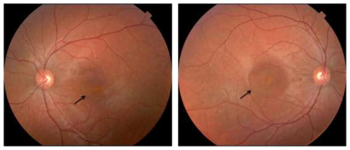
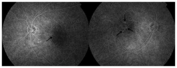
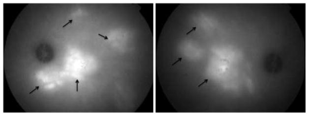
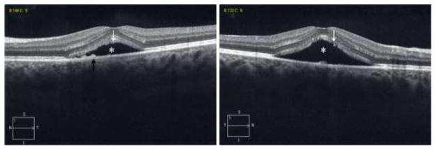
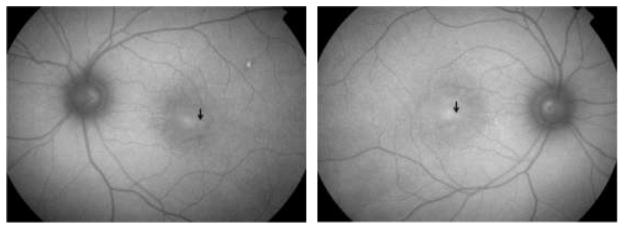
We present a case of bilateral simultaneous central serous chorioretinopathy (CSCR) in a teenage girl with a history of systemic arterial hypertension. A 19-year-old Caucasian female, with a history of systemic arterial hypertension, presented with gradual decrease in her central vision for 1 month. She was diagnosed with bilateral simultaneous CSCR, based on the findings of spectral domain optical coherence tomography (SD-OCT), indocyanine green angiography (ICG), fundus auto-fluorescence, fluorescein angiography and color fundus photographs, which are described. Blood pressure was 134/95 mmHg at presentation. Systemic evaluation failed to reveal a cause for the high blood pressure, and included a panel of blood tests, which were all normal. Her best-corrected visual acuity was 20/30 OD and 20/25 OS. Dilated fundus examination showed normal optic discs and retinal vasculature, with no evidence of hypertensive retinopathy. However, shallow retinal fluid associated with pigmentary changes was noted in the center of both maculae. OCT and ICG findings were consistent with the diagnosis of bilateral CSCR. CSCR can manifest in patients with demographics outside the range of those previously reported. This is the first report of CSCR occurring in a teenage girl, with a history of systemic arterial hypertension. It is important to consider this disease in any patient who has a clinically compatible presentation.
Conflict of interest statement
Figures
References
-
- Yap EY, Robertson DM. The long-term outcome of central serous chorioretinopathy. Arch Ophthalmol. 1996;114:689–692. - PubMed
-
- Gäckle HC, et al. Clinical, fluorescein angiographic and demographic aspectsin central serous chorioretinopathy. Der Ophthalmologe. 1998;95:529–533. - PubMed
-
- Schatz H, Madeira D, Johnson RN, McDonald HR. Central serous chorioretinopathy occurring in patients 60 years of age and older. Ophthalmology. 1992;99:63–67. - PubMed
-
- Spaide RF, Campeas L, Haas A, et al. Central serous chorioretinopathy in younger and older adults. Ophthalmology. 1996;103:2070–2079. - PubMed
-
- Fine SL, Owens SL. Central serous chorioretinopathy in a 7 year-old girl. Am J Ophthalmol. 1980 Dec;90(6):871–3. - PubMed
Publication types
MeSH terms
Grants and funding
LinkOut - more resources
Full Text Sources
Medical
