

Microdomains in forebrain spines: an ultrastructural perspective
- PMID: 22983912
- PMCID: PMC3538892
- DOI: 10.1007/s12035-012-8345-y
Microdomains in forebrain spines: an ultrastructural perspective
Abstract
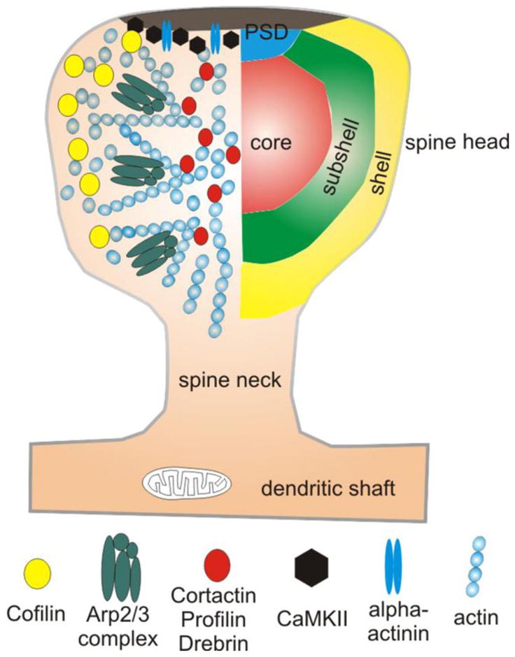
Glutamatergic axons in the mammalian forebrain terminate predominantly onto dendritic spines. Long-term changes in the efficacy of these excitatory synapses are tightly coupled to changes in spine morphology. The reorganization of the actin cytoskeleton underlying this spine "morphing" involves numerous proteins that provide the machinery needed for adaptive cytoskeletal remodeling. Here, we review recent literature addressing the chemical architecture of the spine, focusing mainly on actin-binding proteins (ABPs). Accumulating evidence suggests that ABPs are organized into functionally distinct microdomains within the spine cytoplasm. This functional compartmentalization provides a structural basis for regulation of the spinoskeleton, offering a novel window into mechanisms underlying synaptic plasticity.
Figures
Similar articles
-
The structure and function of actin cytoskeleton in mature glutamatergic dendritic spines.Brain Res. 2014 Jul 21;1573:1-16. doi: 10.1016/j.brainres.2014.05.024. Epub 2014 May 20. Brain Res. 2014. PMID: 24854120 Review.
-
Organization and dynamics of the actin cytoskeleton during dendritic spine morphological remodeling.Cell Mol Life Sci. 2016 Aug;73(16):3053-73. doi: 10.1007/s00018-016-2214-1. Epub 2016 Apr 22. Cell Mol Life Sci. 2016. PMID: 27105623 Free PMC article. Review.
-
Signaling in dendritic spines and spine microdomains.Curr Opin Neurobiol. 2012 Jun;22(3):389-96. doi: 10.1016/j.conb.2012.03.003. Epub 2012 Mar 27. Curr Opin Neurobiol. 2012. PMID: 22459689 Free PMC article. Review.
-
Plasticity of dendritic spines: subcompartmentalization of signaling.Annu Rev Physiol. 2014;76:365-85. doi: 10.1146/annurev-physiol-021113-170400. Epub 2013 Nov 6. Annu Rev Physiol. 2014. PMID: 24215443 Free PMC article. Review.
-
Actin Tyrosine-53-Phosphorylation in Neuronal Maturation and Synaptic Plasticity.J Neurosci. 2016 May 11;36(19):5299-313. doi: 10.1523/JNEUROSCI.2649-15.2016. J Neurosci. 2016. PMID: 27170127 Free PMC article.
Cited by
-
The actin binding protein drebrin helps to protect against the development of seizure-like events in the entorhinal cortex.Sci Rep. 2021 Apr 21;11(1):8662. doi: 10.1038/s41598-021-87967-5. Sci Rep. 2021. PMID: 33883605 Free PMC article.
-
Interrogating Synaptic Architecture: Approaches for Labeling Organelles and Cytoskeleton Components.Front Synaptic Neurosci. 2019 Aug 23;11:23. doi: 10.3389/fnsyn.2019.00023. eCollection 2019. Front Synaptic Neurosci. 2019. PMID: 31507402 Free PMC article. Review.
-
α4βδ-GABAARs in the hippocampal CA1 as a biomarker for resilience to activity-based anorexia.Neuroscience. 2014 Apr 18;265:108-23. doi: 10.1016/j.neuroscience.2014.01.011. Epub 2014 Jan 18. Neuroscience. 2014. PMID: 24444828 Free PMC article.
-
Nanoscale segregation of actin nucleation and elongation factors determines dendritic spine protrusion.EMBO J. 2014 Dec 1;33(23):2745-64. doi: 10.15252/embj.201488837. Epub 2014 Oct 7. EMBO J. 2014. PMID: 25293574 Free PMC article.
-
The Actin Cytoskeleton and Actin-Based Motility.Cold Spring Harb Perspect Biol. 2018 Jan 2;10(1):a018267. doi: 10.1101/cshperspect.a018267. Cold Spring Harb Perspect Biol. 2018. PMID: 29295889 Free PMC article. Review.
References
Publication types
MeSH terms
Grants and funding
LinkOut - more resources
Full Text Sources
