. 2012 Dec;33(12):2441-9.
doi: 10.1093/carcin/bgs286. Epub 2012 Sep 15.

Boswellic acid exerts antitumor effects in colorectal cancer cells by modulating expression of the let-7 and miR-200 microRNA family

Affiliations

Boswellic acid exerts antitumor effects in colorectal cancer cells by modulating expression of the let-7 and miR-200 microRNA family

Masanobu Takahashi et al. Carcinogenesis. 2012 Dec.

Abstract

Colorectal cancer (CRC) is a complex disease with genetic and epigenetic alterations in many key oncogenes and tumor suppressor genes. The active principle of a gum resin from Boswellia serrata, 3-acetyl-11-keto-β-boswellic acid (AKBA), has recently gained attention as a chemopreventive compound due to its ability to target key oncogenic proteins such as 5-lipoxygenase and nuclear factor-kappaB. AKBA has been shown to inhibit the growth of CRC cells; however, the precise molecular mechanisms underlying its anticancer activities in CRC remain unclear. We hypothesized that boswellic acids may achieve their chemopreventive effects by modulating specific microRNA (miRNA) pathways. We found that AKBA significantly up-regulated expression of the let-7 and miR-200 families in various CRC cell lines. Both let-7 and miR-200 are putative tumor-suppressive miRNAs. AKBA modulated the expression of several downstream targets of the let-7 and miR-200 families, such as CDK6, vimentin and E-cadherin. These data were further strengthened by miRNA knockdown studies, which revealed that inhibition of let-7i facilitated enhanced cancer cell proliferation, migration and invasion. In addition, AKBA also induced similar modulation of the let-7 and miR-200 downstream genes in CRC tumors orthotopically implanted in nude mice. These results indicate that AKBA-induced antitumor effects in CRC occur, at least partly through the up-regulation of specific miRNA pathways. Our data provide novel evidence that anticancer effects of boswellic acids are due in part to their ability to regulate cellular epigenetic machinery and further highlight the promise for this phytochemical in the preventative and therapeutic applications of CRC.


Figures
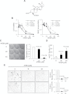
Fig. 1.
Fig. 1.
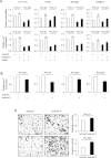
AKBA inhibits cell viability, proliferation, colony formation, migration/invasion and induces apoptosis in CRC cells. (A) Molecular structure of AKBA. (B) Cell viability of four CRC cell lines treated with dimethyl sulfoxide alone or AKBA in a MTT assay (left panel) and a BrdU assay (right panel). Clonogenic survival (C) and apoptotic fractions (D) of HCT116 cells treated with AKBA. (E) Migration (upper panel) and invasion activities (lower panel) of HCT116 cells treated with AKBA. Data are obtained from three or more independent experiments. The P value is calculated by paired or non-paired two-sided Student’s t-test.
Fig. 2.
Fig. 2.
AKBA inhibits NF-κB-related pathways in CRC cells. Western blot analyses for proteins involved in the NF-κB-related pathway in HCT116 cells. AKBA treatment for 24h resulted in inhibition of these proteins in a dose-dependent manner.
Fig. 3.
Fig. 3.
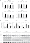
AKBA up-regulates let-7 and miR-200 expression and regulates their downstream targets in CRC cells. (A) The expression of let-7 and miR-200 in four CRC cell lines treated with dimethyl sulfoxide alone or 30 µM of AKBA for 72h. The mRNA (B) and protein expression (C) of the direct or downstream target genes in the cell lines treated with dimethyl sulfoxide alone or 30 µM of AKBA. Data are obtained from three or more independent experiments. The P value is calculated by paired two-sided Student’s t-test. Numbers below each band represent the relative levels of each protein normalized to β-actin, as determined by densitometry.
Fig. 4.
Fig. 4.
let-7i down-regulation causes an increase in cell viability and proliferation, and migration/invasion. (A) Cell viability determined using a MTT assay (upper panel) and cell proliferation using the BrdU incorporation index (lower panel) of the four CRC cell lines treated with negative control inhibitor (anti-NC) or let-7i inhibitor, and subsequently treated with dimethyl sulfoxide alone or 30 µM of AKBA. (B) The expression levels of let-7i in the four cell lines treated with antinegative control inhibitor or let-7i inhibitor. (C) Migration (upper panel) and invasion activity (lower panel) of HCT116 cells transfected with negative control inhibitor or let-7i inhibitor. Data are obtained from three or more independent experiments. The P value is calculated by paired two-sided Student’s t-test.
Fig. 5.
Fig. 5.
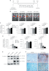
AKBA modulates the expression of let-7 and miR-200 and downstream target genes in orthotopic mice tumors. (A) The schematic diagram of the AKBA treatment protocol in mice. (B) Bioluminescent images of luciferase vector-containing tumors in anesthetized mice. (C) The expression of let-7 and miR-200 in orthotopically implanted tumors treated with vehicle (N = 6) or AKBA with indicated doses (N = 5 per each treatment group). (D) The expression of let-7 and miR-200 target genes (CDK6, E-cadherin and vimentin) in the orthotopic tumors. (E) Western blot analysis of E-cadherin expression in the tumors. (F) A representative of immunohistochemical photomicrograph for E-cadherin staining in vehicle and AKBA-treated (100mg/kg/day) tumors from animals. The P value is calculated by non-paired two-sided Student’s t-test.

Similar articles

Cited by

References

    1. Jemal A., et al. (2010). Cancer statistics, 2010. CA. Cancer J. Clin. 60 277–300 - PubMed
    1. Jemal A., et al. (2011). Global cancer statistics. CA. Cancer J. Clin. 61 69–90 - PubMed
    1. Calin G.A., et al. (2006). MicroRNA signatures in human cancers. Nat. Rev. Cancer 6 857–866 - PubMed
    1. Garzon R., et al. (2009). MicroRNAs in Cancer. Annu. Rev. Med. 60 167–179 - PubMed
    1. Peter M.E. (2009). Let-7 and miR-200 microRNAs: guardians against pluripotency and cancer progression. Cell Cycle 8 843–852 - PMC - PubMed

Publication types