. 2012 Oct 15;189(8):3859-68.
doi: 10.4049/jimmunol.1200940. Epub 2012 Sep 14.

Receptor cross-talk spatially restricts p-ERK during TLR4 stimulation of autoreactive B cells

Affiliations

Receptor cross-talk spatially restricts p-ERK during TLR4 stimulation of autoreactive B cells

Sang-Ryul Lee et al. J Immunol. .

Abstract

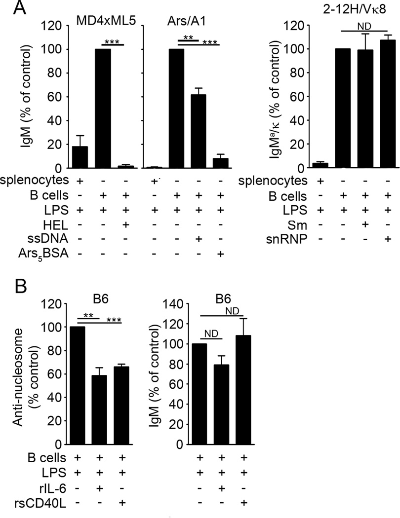
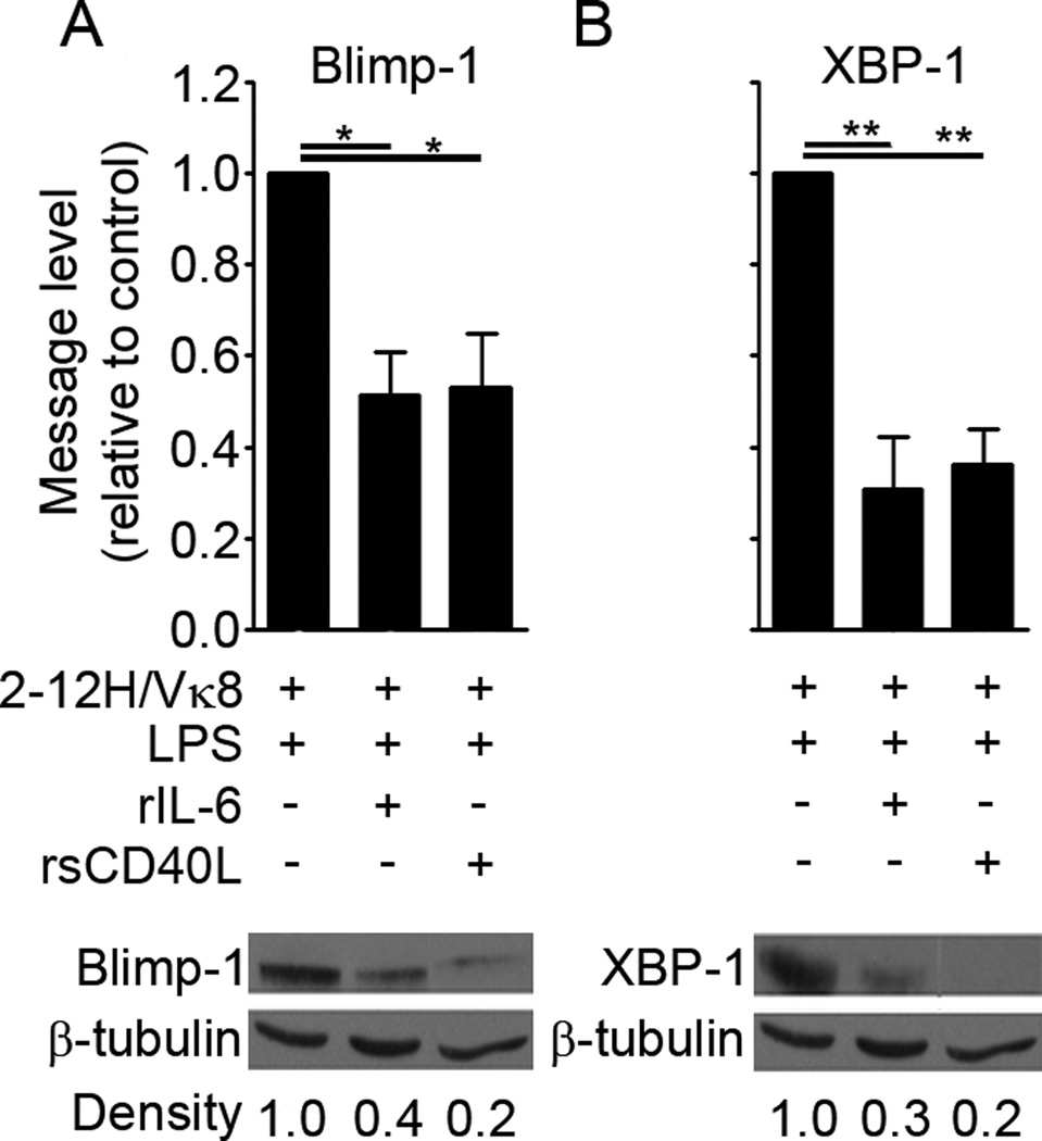
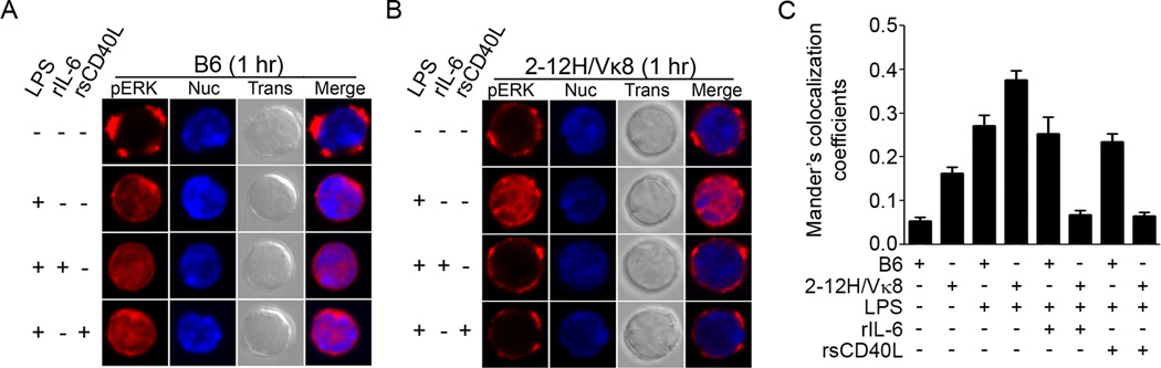
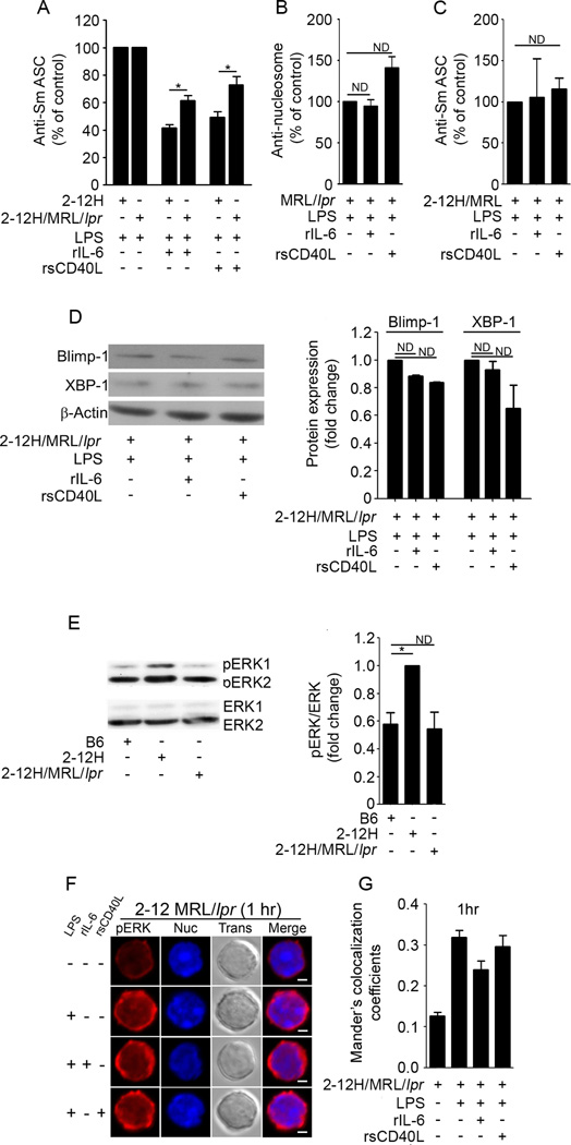
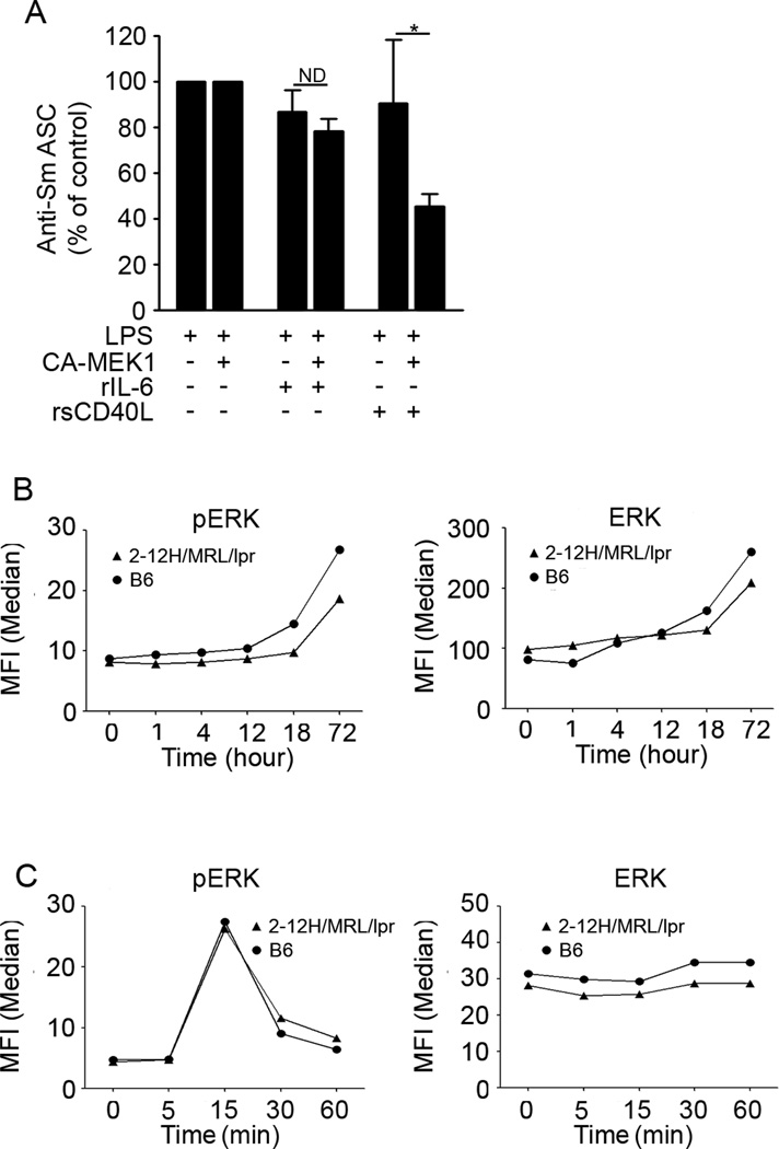
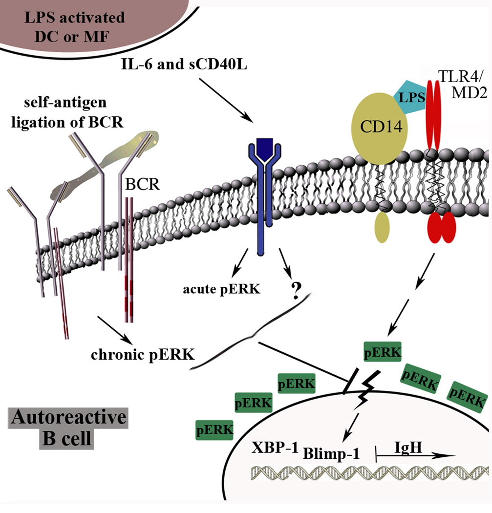
To maintain tolerance, autoreactive B cells must regulate signal transduction from the BCR and TLRs. We recently identified that dendritic cells and macrophages regulate autoreactive cells during TLR4 activation by releasing IL-6 and soluble CD40 ligand (sCD40L). These cytokines selectively repress Ab secretion from autoreactive, but not antigenically naive, B cells. How IL-6 and sCD40L repress autoantibody production is unknown. In this work, we show that IL-6 and sCD40L are required for low-affinity/avidity autoreactive B cells to maintain tolerance through a mechanism involving receptor cross-talk between the BCR, TLR4, and the IL-6R or CD40. We show that acute signaling through IL-6R or CD40 integrates with chronic BCR-mediated ERK activation to restrict p-ERK from the nucleus and represses TLR4-induced Blimp-1 and XBP-1 expression. Tolerance is disrupted in 2-12H/MRL/lpr mice where IL-6 and sCD40L fail to spatially restrict p-ERK and fail to repress TLR4-induced Ig secretion. In the case of CD40, acute signaling in B cells from 2-12H/MRL/lpr mice is intact, but the chronic activation of p-ERK emanating from the BCR is attenuated. Re-establishing chronically active ERK through retroviral expression of constitutively active MEK1 restores tolerance upon sCD40L, but not IL-6, stimulation, indicating that regulation by IL-6 requires another signaling effector. These data define the molecular basis for the regulation of low-affinity autoreactive B cells during TLR4 stimulation; they explain how autoreactive but not naive B cells are repressed by IL-6 and sCD40L; and they identify B cell defects in lupus-prone mice that lead to TLR4-induced autoantibody production.


Conflict of interest statement

Conflict-of-interest disclosure: The authors declare no conflicting financial interests.

Figures

Figure 1
Figure 1. Low-affinity/avidity antigens fail to regulate TLR-induced Ig secretion
Splenic B cells (1 × 10⁵) from MD4 × ML5 (A, left panel), Ars/A1 (A, middle panel), and 2-12H/Vκ8 (A, right panel) mice were stimulated for 4 days with LPS (30 µg/ml) in the absence or presence of HEL (100 µg/ml), ssDNA (500 ng/ml), Ars5BSA (2 µg/ml), Sm (50 U/ml), snRNP (50 µg/ml). IgM or IgMa/κ antibody levels were quantitated by ELISA. In the LPS-stimulated controls, 100% reflects Ig in the range of 8–16 µg/ml (MD4 × ML5), 2–5µg/ml (Ars/A1), and 2–3 µg/ml (2-12H/Vκ8). Splenic B cells (1 × 10⁵) from B6 mice (B) were stimulated for 4 days with LPS (30 µg/ml) in the absence or presence of rIL-6 (30 ng/ml), or rsCD40L (75 ng/ml). Anti-nucleosome Igand total IgM levels were quantitated by ELISA. In the LPS-stimulated controls, 100% reflects 18–42 µg/ml anti-nucleosome Ig, and 14–44 µg/ml total IgM. The data shown represent at least 3 experiments.(*** P<0.001, **P<0.01, ND: no difference)
Figure 2
Figure 2. IL-6 and sCD40L decrease the levels of Blimp-1 and XBP-1
2-12H/Vκ8 B cells were stimulated for 3 days with LPS (30 µg/ml) or LPS combined with rIL-6 (30 ng/ml) or rsCD40L (75 ng/ml). The relative levels of Blimp-1 (A; upper left) and XBP-1 (B; upper right) message were measured by real-time PCR (**P–0.01, *P<0.05). 2-12H/Vκ8 B cells were cultured for 3 days with LPS or LPS combined with rIL-6 or rsCD40L. Lysates from 3 × 10⁶ cells were immunoblotted for Blimp-1 (A; lower left) and XBP-1 (B; lower right).The density of each band was quantitated using Image J, and the fold changes were calculated relative to β-tubulin. The data shown represent at least 3 experiments.
Figure 3
Figure 3. IL-6 and sCD40L repress TLR4-induced Ig in a MEK-dependent manner
3 × 10⁶ purified ex vivo B6 and 2-12H/Vκ8 B cells were lysed and immunoblotted for pERK and total ERK1/2. The density of each band was quantitated with ImageJ, and the ratio of pERK to total ERK1/2 was calculated and expressed as fold change (A). 2-12H/Vκ8 B cells were stimulated for 4 days with LPS or LPS combined with rIL-6 or rsCD40L, in the absence or presence of U0126 (0.5 µM)(B). 3 × 10⁶ splenic B cells from 2-12H/Vκ8 mice were stimulated with LPS (30 µg/ml) in the absence or presence of rIL-6 (30 ng/ml)or rsCD40L (75 ng/ml) for 18–24 hrs followed by DN-MEK1 gene transfer. Two days later, GFP+ B cells (1 × 10⁵) were sorted and stimulated under the same conditions for an additional 48 hrs. IgMa/κ was quantitated by ELISA (C). 1 × 10⁶ splenic B cells from B6 (E and G), or 2-12H/Vκ8 (D and F) mice were stimulated with LPS (30 µg/ml) in the absence or presence of rIL-6 or rsCD40L for various time periods followed by FACS analysis of intracellular pERK and total ERK staining. The data shown represent at least 3 experiments.
Figure 4
Figure 4. IL-6 and sCD40L alter the subcellular localization of pERK in B cells chronically exposed to self-antigen
1 × 10⁶ purified B6 and 2-12H/Vκ8 B cells were stimulated with LPS (30 µg/ml) or LPS in combination with rIL-6 (30 ng/ml) or rsCD40L (75 ng/ml) for 1 hr. Cells were fixed, stained for pERK (red) and dsDNA (blue), and imaged using confocal microscopy (A,B). The co-localization of pERK with the nucleus was quantified in 37–92 cells by calculating the Mander’s coefficient between red and blue fluorescence (Ratio=50%, Threshold Red = 50, Threshold Blue = 30) (C).
Figure 5
Figure 5. IL-6 and sCD40L fail to repress B cells from autoimmune-prone mice
1 × 10⁵ splenic B cells from 2-12H and 2-12H/MRL/lpr mice (5–9 weeks old) were stimulated with LPS (30 µg/ml) in the absence or presence of rIL-6 (30ng/ml) orrsCD40L (75 ng/ml) for 4 days. ASCs were enumerated using ELISPOT assays (A). 1 × 10⁵ splenic B cells from MRL/lpr mice (5–10 weeks old) were stimulated with LPS in the absence or presence of rIL-6 orrsCD40L for 4days. Anti-nucleosome Ig secretion was quantitated by ELISA (B). 1 × 10⁵ splenic B cells from 2-12H/MRL/MpJ (2-12H/MRL)mice (5 –9 weeks old) were stimulated with LPS in the absence or presence of rIL-6 orrsCD40L for 4days. The number of Sm secreting cells was quantitated by ELISPOT (C). 2-12H/MRL/lpr B cells were stimulated for 3 days with LPS (30 µg/ml) in the absence or presence of rIL-6 (30 ng/ml), or rsCD40L (75 ng/ml). Lysates from 3 × 10⁶ cells were immunoblotted for Blimp-1 and XBP-1. The density of each band was quantitated with ImageJ, and the fold change was calculated relative to β-actin (D). 3 × 10⁶ purified B6, 2-12H/Vκ8,and 2-12H/MRL/lpr B cells were lysed and immunoblotted for pERK and total ERK1/2. The density of each band was quantitated with ImageJ and the ratio of pERK/ERK from 3 experiments was calculated (E). 1 × 10⁶ purified ex vivo2-12H/MRL/lpr B cells were stimulated with LPS in the absence or presence of rIL-6 orrsCD40L for 1 hr. Cells were fixed, stained for pERK (red) and dsDNA (blue), and imaged using confocal microscopy (F). Colocalization of pERK with the nucleus at the 1 hr time point was quantified in 40 cells by calculating the Mander’s coefficient between red and blue fluorescence (Ratio=50%, Threshold Red = 50, Threshold Blue = 30) (G). The data shown represent at least 3 experiments.
Figure 6
Figure 6. Expression of constitutively active MEK1 restores the ability of sCD40L to repress TLR4-induced Ig secretion
3 × 10⁶ splenic B cells from 2-12H/MRL/lprmice were stimulated with LPS (30 µg/ml) in the absence or presence of rIL-6 (30 ng/ml)or rsCD40L (75 ng/ml) for 18 – 24 hr followed by CA-MEK1 gene transfer. On day 4, GFP+ B cells were sorted and ASCs were enumerated using ELISPOT assays. Data represent at least 3 experiments. (*P<0.05; ND not different). 1 × 10⁶ splenic B cells from B6 or 2-12H/MRL/lpr mice were stimulated with LPS for various time periods followed by intracellular staining with pERK (B, left panel) and total ERK1/2 (B, right panel). 1 × 10⁶ splenic B cells from B6, and 2-12H/MRL/lpr mice were stimulated with rsCD40L (75 ng/ml) for various time periods followed by FACS analysis of intracellular pERK (C, left panel)and total ERK staining (C, right panel).
Figure 7
Figure 7. Receptor cross-talk regulates autoreactive B cells during TLR4 stimulation
We propose a model wherein the chronic ligation of self-antigen by the BCR distinguishes autoreactive B cells from antigenically naïve cells. Chronic BCR ligation induces chronically active ERK (Figure 3A). In addition to stimulating B cells, LPS induces DCs and MFs to secrete IL-6 and sCD40L, which in turn acutely stimulateIL-6 receptor or CD40 on B cells inducing an acute pERK signal (Figure 6B for CD40). The combined chronic and acute ERK signals exclude pERK from the nucleus, limiting expression of TLR4-induced Blimp-1 and XBP-1 and thereby repressing plasma cell differentiation. Stimulation through IL-6 receptor relies on pERK (Figure 3), but pERK is not sufficient (Figure 6). The identity of the second effector coupled to IL-6 receptor remains unknown, as denoted by the question mark.

Similar articles

Cited by

References

    1. Cambier JC, Gauld SB, Merrell KT, Vilen BJ. B-cell anergy: from transgenic models to naturally occurring anergic B cells? Nat Rev Immunol. 2007;7:633–643. - PMC - PubMed
    1. Luning Prak ET, Monestier M, Eisenberg RA. B cell receptor editing in tolerance and autoimmunity. Ann N Y Acad Sci. 2011;1217:96–121. - PMC - PubMed
    1. Pillai S, Mattoo H, Cariappa A. B cells and autoimmunity. Curr Opin Immunol. 2011;23:721–731. - PMC - PubMed
    1. Ferry H, Leung JC, Lewis G, Nijnik A, Silver K, Lambe T, et al. B-cell tolerance. Transplantation. 2006;81:308–315. - PubMed
    1. Kilmon MA, Rutan JA, Clarke SH, Vilen BJ. Low-affinity, Smith antigen-specific B cells are tolerized by dendritic cells and macrophages. J Immunol. 2005;175:37–41. - PMC - PubMed

Publication types

MeSH terms