Review
. 2013 Jan;48(1):1-9.
doi: 10.1165/rcmb.2012-0282TR. Epub 2012 Sep 13.

Regulation and functional significance of autophagy in respiratory cell biology and disease

Affiliations
Review

Regulation and functional significance of autophagy in respiratory cell biology and disease

Avignat S Patel et al. Am J Respir Cell Mol Biol. 2013 Jan.

Abstract

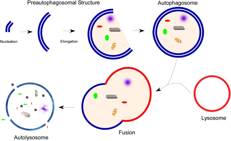
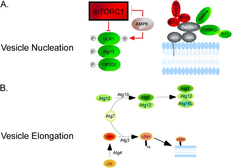
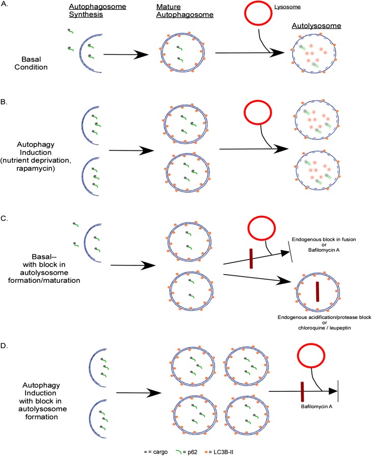
Autophagy is a homeostatic process common to all eukaryotic cells that serves to degrade intracellular components. Among three classes of autophagy, macroautophagy is best understood, and is the subject of this Review. The function of autophagy is multifaceted, and includes removal of long-lived proteins and damaged or unneeded organelles, recycling of intracellular components for nutrients, and defense against pathogens. This process has been extensively studied in yeast, and understanding of its functional significance in human disease is also increasing. This Review explores the basic machinery and regulation of autophagy in mammalian systems, methods employed to measure autophagic activity, and then focuses on recent discoveries about the functional significance of autophagy in respiratory diseases, including chronic obstructive pulmonary disease, cystic fibrosis, tuberculosis, idiopathic pulmonary fibrosis, pulmonary arterial hypertension, acute lung injury, and lymphangioleiomyomatosis.


Figures

Figure 1.
Figure 1.
Overview of macroautophagy. Macroautophagy (autophagy) begins with the nucleation and elongation of a preautophagosomal structure. This structure begins to encompass the cargo intended for degradation, followed by completion of the double-membraned autophagosome. The autophagosome fuses with a lysosome, forming the autolysosome. Lysosomal hydrolases then degrade the contents, which are recycled back to the cytosol through membrane permeases.
Figure 2.
Figure 2.
Molecular machinery of vesicle nucleation and elongation. (A) Vesicle nucleation begins with mammalian target of rapamycin complex (mTORC) 1 down-regulation, which can be affected by AMP-activated protein kinase (AMPK) via TSC2. This results in UNC-51–like kinase (ULK) 1 dephosphorylation and subsequent phosphorylation of Fak-family kinase–interacting protein (FIP)-200 and Atg13. AMPK can also activate ULK1 directly by phosphorylation at a different site than mTORC1. Nucleation also involves the phosphatidylinositol-3-kinase (PI3K) complex, which includes Vps34, Beclin-1, Atg14, and p150/Vps15. Beclin-1 potentiates Vps34 activity. Beclin-1 activity can be modulated through several binding partners: Ambra-1, ultraviolet (UV) radiation resistance–associated gene (UVRAG), and bif-1 binding induces autophagy; Bcl-2 binding prevents autophagosome initiation. The latter interaction is enhanced by inositol 1,4,5-triphosphate receptor (IP3R), nutrient deprivation–autophagy factor-1 (NAF-1). (B) Vesicle elongation involves two ubiquitin-like reactions. Through the action of Atg7 and Atg10, Atg12 is covalently bound to Atg5, forming Atg12-Atg5. This complex conjugates with Atg16L1, forming Atg12-Atg5-Atg16L1. The second reaction revolves around microtubule-associated protein 1 light chain 3/Atg8 (LC3). Atg4B cleaves the LC3 precursor to form cytosolic LC3-I. Atg7 and Atg3 lipidate LC3-I with phosphatidylethanolamine (PE) to form LC3-II, which is targeted to the forming autophagosome.
Figure 3.
Figure 3.
Measurement of autophagic flux. (A) Under basal conditions, there is a low level of autophagosome formation and maturation into an autolysosome. p62 and its cargo, as well as LC3-II along the inner membrane, is degraded in the autolysosome. (B) When autophagy is induced, there is increased autophagosome formation and completion of the pathway, resulting in increased autolyosomes. (C) Under basal conditions, there is a low level of autophagosome formation. However, in the presence of downstream blocks to autolysosome formation or function, autophagosome numbers are increased. p62 does not undergo degradation, and accumulates in this condition. The block can occur endogenously or can be introduced with chemical inhibitors. Bafilomycin A prevents autophagosome and lysosome fusion, chloroquine prevents autolysosomal acidification, and leupeptin inhibits lysosomal protease function. (D) To elucidate whether increased numbers of autophagosomes are due to autophagy induction (B) or downstream block (C), a chemical inhibitor is employed. In this case, introducing bafilomycin to a system where autophagy is induced results in increased numbers of autophagosomes (compared with [B]).

Similar articles

Cited by

References

    1. Tolkovsky AM. Mitophagy. Biochim Biophys Acta 2009;1793:1508–1515 - PubMed
    1. Bernales S, Schuck S, Walter P. ER-phagy: selective autophagy of the endoplasmic reticulum. Autophagy 2007;3:285–287 - PubMed
    1. Sakai Y, Oku M, van der Klei IJ, Kiel JA. Pexophagy: autophagic degradation of peroxisomes. Biochim Biophys Acta 2006;1763:1767–1775 - PubMed
    1. Kraft C, Deplazes A, Sohrmann M, Peter M. Mature ribosomes are selectively degraded upon starvation by an autophagy pathway requiring the UBP3P/BRE5P ubiquitin protease. Nat Cell Biol 2008;10:602–610 - PubMed
    1. Levine B. Eating oneself and uninvited guests: autophagy-related pathways in cellular defense. Cell 2005;120:159–162 - PubMed

Publication types

MeSH terms

LinkOut - more resources