. 2012;7(9):e43463.
doi: 10.1371/journal.pone.0043463. Epub 2012 Sep 12.

Association of shank 1A scaffolding protein with cone photoreceptor terminals in the mammalian retina

Affiliations

Association of shank 1A scaffolding protein with cone photoreceptor terminals in the mammalian retina

Salvatore L Stella Jr et al. PLoS One. 2012.

Abstract

Photoreceptor terminals contain post-synaptic density (PSD) proteins e.g., PSD-95/PSD-93, but their role at photoreceptor synapses is not known. PSDs are generally restricted to post-synaptic boutons in central neurons and form scaffolding with multiple proteins that have structural and functional roles in neuronal signaling. The Shank family of proteins (Shank 1-3) functions as putative anchoring proteins for PSDs and is involved in the organization of cytoskeletal/signaling complexes in neurons. Specifically, Shank 1 is restricted to neurons and interacts with both receptors and signaling molecules at central neurons to regulate plasticity. However, it is not known whether Shank 1 is expressed at photoreceptor terminals. In this study we have investigated Shank 1A localization in the outer retina at photoreceptor terminals. We find that Shank 1A is expressed presynaptically in cone pedicles, but not rod spherules, and it is absent from mice in which the Shank 1 gene is deleted. Shank 1A co-localizes with PSD-95, peanut agglutinin, a marker of cone terminals, and glycogen phosphorylase, a cone specific marker. These findings provide convincing evidence for Shank 1A expression in both the inner and outer plexiform layers, and indicate a potential role for PSD-95/Shank 1 complexes at cone synapses in the outer retina.


Conflict of interest statement

Competing Interests: The authors have declared that no competing interests exist.

Figures

Figure 1
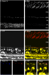
Figure 1. Shank 1A immunoreactivity is in both the inner plexiform layer (IPL) and outer plexiform layer (OPL) of the mouse YFP-16 line retina.
A–C: A. Image of a retinal section immunostained for Shank 1A. B. Mouse YFP-16 line vertical retinal section. C. Shank 1A (red) immunolabeling and YFP (yellow). Shank1A expression is restricted to the OPL and IPL. A regular pattern of Shank 1A immunolabeling appears in the OPL, which is indicative of cone photoreceptor terminals. D–E: High magnification zoom of the OPL demonstrates that Shank 1A puncta (red) are distal to the dendrite tips (yellow) of YFP labeled cone bipolar cells, suggesting that Shank 1A is expressed presynaptic to the YFP cone bipolar cell dendrite. G–L: High magnification zoom of the IPL demonstrates that Shank 1A puncta are likely expressed postsynaptically to bipolar cell terminals. G. Shank 1A immunoreactive puncta. H. YFP labeled neurons and processes within the IPL region. I. PKCα labeled rod bipolar cell axons and terminals. J. Combined Shank 1A (red) and PKCα (blue) immunolabeling illustrate that shank 1A puncta are postsynaptic to rod bipolar cell terminals in the IPL. K. Combined Shank 1A (red) immunolabeling and YFP (yellow) in the IPL demonstrate that Shank 1A puncta are postsynaptic to cone bipolar cell terminals in the IPL. L. Combined triple fluorescent image of Shank 1A (red), PKCα (blue), and YFP (yellow) in the IPL. OPL = outer plexiform layer, INL = inner nuclear layer, IPL = inner plexiform layer, and GCL = ganglion cell layer. Scale bars = 10 µm.
Figure 2
Figure 2. Shank 1A immunoreactivity is absent when the Shank 1 gene has been deleted in mouse retina.
A: Shank 1A immunoreactivity in wild type mouse retina, punctate antibody labeling in the OPL in the outer retina and throughout the IPL. B: No Shank 1A immunoreactivity was found in retinal sections obtained from animals where the Shank 1 gene was deleted. (OS = outer segment, ONL = outer nuclear layer, OPL = outer plexiform layer, INL = inner nuclear layer, IPL = inner plexiform layer, and GCL = ganglion cell layer). Scale bar is 10 µm.
Figure 3
Figure 3. Shank 1A is expressed with PSD-95 labeled photoreceptor terminals in the mouse thy-1.2 YFP 16 line retina.
A–D: Zoomed confocal region of the OPL immunostained with rabbit anti-Shank1A and mouse anti-PSD-95 antibodies. A. Shank 1A puncta labeling in the OPL in the mouse retina. B. PSD-95 immunolabels rod and cone photoreceptor terminals in the OPL. C. YFP labeled dendrites from cone bipolar cells in the OPL. D. Merged confocal image showing Shank 1A (red), PSD-95 (blue), and YFP (yellow). E–J: High magnification single plane confocal images of Shank 1A and PSD-95 in the mouse thy 1.2 YFP 16 line retina (100× objective, N.A. 1.3). E. Shank1 A (red). F. PSD-95 (blue). G. YFP (yellow). H. Merged image of YFP and Shank 1A, YFP and Shank 1A puncta are not co-localized at cone bipolar cell dendrites (see arrowheads, shank fluorescence above YFP dendrite. I. Merged image of Shank 1A and PSD-95, the two immunoreactivities (pink) indicate co-localization of Shank 1a and PSD-95 (see arrows which indicate that Shank 1A is co-localized with PSD-95). Arrows indicate location of Shank 1A immunoractive puncta co-localized with PSD-95. J. Merged image of inset Shank 1A (red), PSD-95 (blue), and YFP (yellow). Primary antibodies were detected using a secondary goat anti-rabbit Alexa 568 IgG for Shank 1A, and a goat anti-mouse Alexa 633 IgG to PSD-95. Scale bar is 5 µm.
Figure 4
Figure 4. Shank 1A immunoreactivity is not associated with the synaptic ribbon protein.
High magnification zoom of a region in the OPL. A–H: Confocal images from the mouse thy-1.2 YFP 16 line immunolabeled with Shank 1A and CtBP2 (a homologue of RIBEYE), a marker of synaptic ribbons in mammalian retina. A. Shank 1A (red). B. CtBP2 (blue). C. A combined fluorescence image showing Shank 1A (red) and CtBP2 (blue). D. A schematic illustrating the zoomed confocal image in H, showing that the Shank 1A puncta (red) is located distal and adjacent to the tips of the YFP dendrite (yellow), and are surrounded by CtBP2 labeled synaptic ribbon protein structures (blue). E. YFP (yellow) F. A combined fluorescence image showing Shank 1A (red) and YFP (yellow). G. A combined fluorescence image showing CtBP2 (blue) and YFP (yellow). H. A combined triple labeled image showing Shank 1A (red), CtBP2 (blue), and YFP (yellow). Scale bar = 10 µm.
Figure 5
Figure 5. Shank 1A immunoreactivity is co-localized with the lectin PNA (peanut agglutinin) in photoreceptor terminals.
A–D: Combined labeling of Shank 1A, PNA, and YFP in the mouse thy-1.2 YFP 16 line vertical retinal section. A. Shank 1A labels both the OPL and IPL. The immunoreactive puncta in the OPL are indicative of cone photoreceptor labeling. B. PNA conjugated rhodamine labels the inner and outer segments of cone photoreceptors, and cone photoreceptor terminals in the OPL. C. YFP fluorescence is present in bipolar, amacrine, and ganglion cells. D. Combined triple fluorescence channel image of PNA (red), Shank 1A (blue), and YFP (yellow). A box is drawn of a region in the OPL and high magnification images are shown in E–H. E–H: High magnification zoom of a region in the OPL from D. E. Shank 1A (blue). F. PNA (red). G. YFP labeled cone bipolar cells and their dendrites (yellow). H. A combined triple labeled fluorescent image showing that Shank 1A (blue) is expressed at the same site as PNA (red) above the YFP cone bipolar cell dendrites (yellow). OS = outer segment, IS = inner segment, ONL = outer nuclear layer, OPL = outer plexiform layer, INL = inner nuclear layer, IPL = inner plexiform layer, and GCL = ganglion cell layer. Scale bars is 10 µm.
Figure 6
Figure 6. Shank 1A immunoreactivity co-localizes with the lectin WGA at cone photoreceptor terminals.
A–C: A. Shank 1A B. WGA labeling at the OPL. Arrowheads indicate WGA rod spherule labeling, and arrows indicate the location of WGA labeling of cone pedicles. C. YFP cone bipolar cell dendrites. D–F: Shank 1A (blue) and WGA (red) co-localize at cone terminals in the OPL. G–I: YFP cone bipolar cell dendrites (yellow) synapse with WGA (red) labeled cone terminals, which cradle Shank 1A immunoreactive puncta (blue). Scale bar is 10 µm.
Figure 7
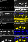
Figure 7. Shank 1 immunoreactivity is expressed solely within cone terminals of the mouse thy-1.2 YFP 16 line retina.
A–D: Shank 1A is co-localized with glycogen phsophorylase (cone photoreceptor marker). A–D: Combined labeling of Shank 1A, glycogen phosphorylase, and YFP in a vertical retinal section. A. Shank 1A labels both the OPL and IPL. The immunoreactive puncta in the OPL are indicative of cone photoreceptor labeling. B. YFP fluorescence is present in bipolar, amacrine, and ganglion cells. C. Glycogen phosphorylase (GP), a cone photoreceptor marker strongly labels the cone terminals with faint labeling of bipolar cell bodies and their axons. D. Combined triple label fluorescence image of Shank 1A (red), YFP (yellow), and glycogen phosphorylase (GP) (blue), and. A box is drawn of a region in the OPL and high magnification images are shown in E–J. E–J: High magnification zoom of a region in the OPL from the inset above in Fig. 7D. E. Shank 1A. F. Merge of Shank 1A (red) and YFP (yellow), showing that Shank 1A (red) is expressed above the cone bipolar cell dendrites (yellow). G. Glycogen phosphorylase (GP). H. Merge of Shank 1A (red) and Glycogen phosphorylase (GP), the two immunoreactivities (pink) indicate co-localization of Shank 1a and Glycogenphosphorylase (GP). I. YFP labeled cone bipolar cells and their dendrites. J. Combined image of Shank 1A (red), glycogen phosphorylase (blue), and YFP cone bipolar cell dendrites (yellow). OPL = outer plexiform layer, INL = inner nuclear layer, IPL = inner plexiform layer, and GCL = ganglion cell layer. Scale bar is 10 µm.

References

    1. Koulen P, Fletcher EL, Craven SE, Bredt DS, Wässle H (1998) Immunocytochemical localization of the postsynaptic density protein PSD-95 in the mammalian retina. J Neurosci 18: 10136–10149. - PMC - PubMed
    1. Kennedy MB (1997) The postsynaptic density at glutamatergic synapses. Trends Neurosci 20: 264–268. - PubMed
    1. Sheng M (2001) The postsynaptic NMDA-receptor–PSD-95 signaling complex in excitatory synapses of the brain. J Cell Sci 114: 1251. - PubMed
    1. Olson PA, Tkatch T, Hernandez-Lopez S, Ulrich S, Ilijic E, et al. (2005) G-protein-coupled receptor modulation of striatal CaV1.3 L-type Ca2+ channels is dependent on a Shank-binding domain. J Neurosci 25: 1050–1062. - PMC - PubMed
    1. Sala C, Futai K, Yamamoto K, Worley PF, Hayashi Y, et al. (2003) Inhibition of dendritic spine morphogenesis and synaptic transmission by activity-inducible protein Homer1a. J Neurosci 23: 6327–6337. - PMC - PubMed

Publication types

LinkOut - more resources