. 2012;7(9):e43783.
doi: 10.1371/journal.pone.0043783. Epub 2012 Sep 11.

A detailed, hierarchical study of Giardia lamblia's ventral disc reveals novel microtubule-associated protein complexes

Affiliations

A detailed, hierarchical study of Giardia lamblia's ventral disc reveals novel microtubule-associated protein complexes

Cindi L Schwartz et al. PLoS One. 2012.

Abstract

Giardia lamblia is a flagellated, unicellular parasite of mammals infecting over one billion people worldwide. Giardia's two-stage life cycle includes a motile trophozoite stage that colonizes the host small intestine and an infectious cyst form that can persist in the environment. Similar to many eukaryotic cells, Giardia contains several complex microtubule arrays that are involved in motility, chromosome segregation, organelle transport, maintenance of cell shape and transformation between the two life cycle stages. Giardia trophozoites also possess a unique spiral microtubule array, the ventral disc, made of approximately 50 parallel microtubules and associated microribbons, as well as a variety of associated proteins. The ventral disc maintains trophozoite attachment to the host intestinal epithelium. With the help of a combined SEM/microtome based slice and view method called 3View® (Gatan Inc., Pleasanton, CA), we present an entire trophozoite cell reconstruction and describe the arrangement of the major cytoskeletal elements. To aid in future analyses of disc-mediated attachment, we used electron-tomography of freeze-substituted, plastic-embedded trophozoites to explore the detailed architecture of ventral disc microtubules and their associated components. Lastly, we examined the disc microtubule array in three dimensions in unprecedented detail using cryo-electron tomography combined with internal sub-tomogram volume averaging of repetitive domains. We discovered details of protein complexes stabilizing microtubules by attachment to their inner and outer wall. A unique tri-laminar microribbon structure is attached vertically to the disc microtubules and is connected to neighboring microribbons via crossbridges. This work provides novel insight into the structure of the ventral disc microtubules, microribbons and associated proteins. Knowledge of the components comprising these structures and their three-dimensional organization is crucial toward understanding how attachment via the ventral disc occurs in vivo.


Conflict of interest statement

Competing Interests: The authors have declared that no competing interests exist.

Figures

Figure 1
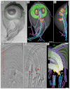
Figure 1. The complex microtubule cytoskeleton of Giardia reconstructed by 3View® and plastic-section tomography.
A) Selected SEM slice (back-scattered electron signal) showing eight flagella [anterior flagella (AFL); caudal flagella (CFL); posterior-lateral flagella (PFL); and ventral flagella (VFL)], part of the ventral disc (VD: green outline), the bare area (BA), the lateral shield (LS), and lateral crest (LC). B) 3-D model of a whole-cell reconstruction: ventral disc, nucleus (N), median body (MB), and the four pairs of flagella. C) The side-view of the model shows that the entire microtubule cytoskeleton is located in the ventral part of the cell. D) 5 nm tomographic slice from a montaged, plastic serial section tomogram of a portion of the ventral disc. At the most ventral part of the disc, there are parallel microtubules and microribbons. The relationship of the disc to the helical axis is as indicated: margin-facing (M) or axis-facing (A). The bare area (BA) is also indicated. E) 5 nm tomographic slice showing the arrangement of four basal bodies and how the microtubules (MT) of the ventral disc originate from dense bands (arrows). F) Model from the tomographic reconstruction showing the supernumerary microtubules (yellow) are ventral to the ventral disc microtubules (white). Microtubule ends are classified as either capped (red dots, arrows) or open (green dots). Microribbons are shown in green. One of the anterior flagella (purple) penetrates the overlap zone. Scale bars in A–C = 2 µm, D–F = 200 nm.
Figure 2
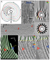
Figure 2. Cryo-electron tomography of the ventral disc.
To describe the orientation, the dorsal-ventral (V) line and the axis-facing and margin-facing (M) sides of the microtubule are labeled. Microtubule polarity is indicated (+). A) 75 nm cryo-tomographic XZ-slice from Tomo-1 showing the ventral disc, an array of parallel microtubules with microribbons. The power spectrum (inset) shows the regular ∼55 nm spacing between neighboring microtubule-microribbon complexes. The missing wedge, a limitation inherited to tomographic data, is illustrated by the large, black, wedge-shaped area in the power spectrum. B) 25 nm tomographic XY-slice from box B in panel A. The power spectrum (inset) shows a repetitive unit every 8 nm. C) 25 nm tomographic XY-slice from box C in panel A. Microribbons have 16 nm repeats (power spectrum, inset). The lateral spacing of the microribbons (and underlying microtubules) is much closer at the margin of the disc (red lines). Scale bars, 50 nm.
Figure 3
Figure 3. Polarity of ventral disc microtubules is unambiguous with minus-ends originating at dense bands or at the inside edge of the spiral.
A) Sketch showing the ventral disc with the dorsal side pointing torward the viewer. Microtubules start with their minus-ends near the overlap zone and spiral downward to the ventral side, thereby forming a left-handed helix. The repetitive units are on the margin-facing side (→M) of the microtubules (dotted colored lines). B) A 10 nm plastic-tomographic slice shows capped microtubule ends (blue arrows) at the dense bands, indicating their minus-ends, while panel C) shows open microtubule ends (red arrows) at the periphery of the disc, typical for plus-ends. D) An end-on view from a helical reconstruction of a bovine microtubule decorated with kinesin-1 motor domains (K) (for example see [61]); when viewed from the minus-end, tubulin (T) shows a right-handed slew while globular microtubule-associated proteins (MAPs) such as kinesin motor domains often bend torward the left (lower panel). The same pattern is visible in the 3-D average of the tomographic reconstruction (upper panel), though with less clarity due to the missing wedge effects. E) A portion of the model of the plastic-section tomogram from Figure 1D showing a plus-end (red arrow) of a microtubule that is ending within the spiral and a minus-end (blue arrow) beginning at the inner edge of the spiral. The microribbons (green) of the inserted microtubules (white) are proximal to the minus-end (blue arrow) of the microtubule. F, G) The upper panels show the plastic-section tomogram in cross-section with the microribbons modeled in green and microtubules in white. The yellow line shows the line of rotation 90° to make the views in the lower panels. The microtubule ending within the spiral (red arrow) is slightly below the neighboring microtubules. The microtubule beginning at the inner edge of the spiral (blue arrow) starts above the neighboring microtubules. Scale bars in B and C = 50 nm; D = 5 nm; E = 50 nm; F and G = 25 nm. Panel A: adapted with permission from .
Figure 4
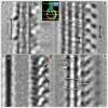
Figure 4. Grand average of the ventral disc microtubule-microribbon complex.
Selected 0.776 nm XY-slices from the grand average (inset shows the location of each slice of the grand average). A) Microribbons are made of three sheets; axis-facing (A), inner sheet (I), and margin-facing (M). The overall microribbon structure shows a distinct 16 nm repeat over two successive αβ-tubulin dimers. Crossbridges (CB) are best visible on the margin-facing sheet but most likely contact the axis-facing sheet on the other side. B) Slice through the dorsal-facing microtubule wall near the microribbon attachment point showing the α- and β-tubulin densities and the regions of the side-arms (SA) forming an axial 8 nm repeat. The white line shows the characteristic pseudo-helix of neighboring protofilaments (PF6–9), which are composed of α- and β-tubulin subunits at a 4 nm spacing. C) The lumen (L) of the microtubule is empty. The associated side-arms are connected laterally (arrow). D) A slice through the most ventral microtubule-associated densities (gMAP1, gMAP2, and P) reveals the position of the microtubule seam—the offset is between gMAP1 and the paddle (P), but there is no offset between gMAP2 and gMAP1. Scale bar = 5 nm.
Figure 5
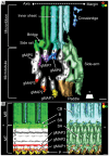
Figure 5. Isosurface representation of the grand average.
A) View along the microtubule axis torward the plus-end. Each protofilament is numbered clockwise, starting with 1 at the location of the seam (see Figure 4D). Microribbons consist of three parallel sheets: axis-facing sheet, inner sheet, and margin-facing sheet. The crossbridges are visible on the margin-facing sheet. The axis-facing sheet is connected to the side rail, a fibrous structure attached to protofilament 6, via the bridge. There are several giardial microtubule inner proteins (gMIPs) associated with the inner wall of the microtubule on protofilaments 5, 7, and 8 (gMIP5, gMIP7, and gMIP8). There are also giardial microtubule-associated proteins (gMAPs) attached to the outer wall at protofilaments 1, 2, and 3 (gMAP1, gMAP2, and gMAP3). The side-arms (SA) span protofilaments 9–12 and are associated with the paddle (P), which is connected to protofilament 13. B) Axis-facing (left) and margin-facing (right) views. The axis-facing side has 2 “naked” protofilaments (PF4 and PF5). All three gMAPs (gMAP1, gMAP2, gMAP3) are visible as well as part of the paddle (P). The side rail (SR) on protofilament 6 is connected to the axis-facing sheet of the microribbon via the bridge (B). On the margin-facing side, gMAP1 is barely visible behind the paddle. The side-arm covers the rest of the microtubule. The crossbridges (CB) are evident on the margin-facing sheet (M) of the microribbon. Scale bars = 5 nm.
Figure 6
Figure 6. MIPs on the microtubule inner side of the grand average.
A) Isosurface representation of the inner microtubule wall and associated gMIPs. While gMIP5 and gMIP7 appear regularly every 8 nm, according to the αβ-tubulin dimer repeat, gMIP8 clearly exhibits a 16 nm repeat that reaches over two consecutive dimers along protofilament 8. Panels B and C show vertical 0.776 nm slices through the volume in A, cutting through protofilament 7 (B) and protofilament 8 (C), respectively. Scale bars = 10 nm.
Figure 7
Figure 7. Microribbons of the ventral disc are composed of three parallel sheets.
A) The bridge (B) connects the axis-facing sheet (A) to the side rail (SR). In addition, there is a complex array of proteins connecting the side-arm (SA) to the margin-facing sheet (M). The inner sheet (I) of the microribbon is partially associated with the margin-facing sheet. B) Lateral connections between the three sheets are evident (arrowheads). C) The axis-facing sheet has a distinct 16 nm repeat of 3 domains, one of which sticks out and is likely the attachment site of the crossbridges (CB) from the neighboring microribbon. D) A faint 5 nm repeat is evident along the dorsal-ventral line of the axis-facing sheet. E) The grand average isosurface has been inserted back into the original data, where each segment of the model has been rotated and shifted according to the parameters derived from the average. Dotted red lines have been drawn to indicate that the crossbridges on the margin-facing side likely connect to the 16 nm protrusions on the axis-facing side of the microribbon. Scale bars A–D, schematic = 5 nm, E = 25 nm.

Similar articles

Cited by

References

    1. Savioli L, Smith H, Thompson A (2006) Giardia and Cryptosporidium join the “Neglected Diseases Initiative”. Trends in parasitology 22: 203–208. - PubMed
    1. Thompson RC (2000) Giardiasis as a re-emerging infectious disease and its zoonotic potential. International journal for parasitology 30: 1259–1267. - PubMed
    1. Adam RD (2001) Biology of Giardia lamblia. Clinical microbiology reviews 14: 447–475. - PMC - PubMed
    1. Land KM, Johnson PJ (1999) Molecular basis of metronidazole resistance in pathogenic bacteria and protozoa. Drug resistance updates: reviews and commentaries in antimicrobial and anticancer chemotherapy 2: 289–294. - PubMed
    1. Cyrklaff M, Kudryashev M, Leis A, Leonard K, Baumeister W, et al. (2007) Cryoelectron tomography reveals periodic material at the inner side of subpellicular microtubules in apicomplexan parasites. The Journal of experimental medicine 204: 1281–1287. - PMC - PubMed

Publication types

MeSH terms