. 2012;7(9):e44263.
doi: 10.1371/journal.pone.0044263. Epub 2012 Sep 12.

Polar/Ionizable residues in transmembrane segments: effects on helix-helix packing

Affiliations

Polar/Ionizable residues in transmembrane segments: effects on helix-helix packing

Manuel Bañó-Polo et al. PLoS One. 2012.

Abstract

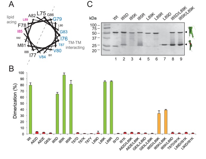
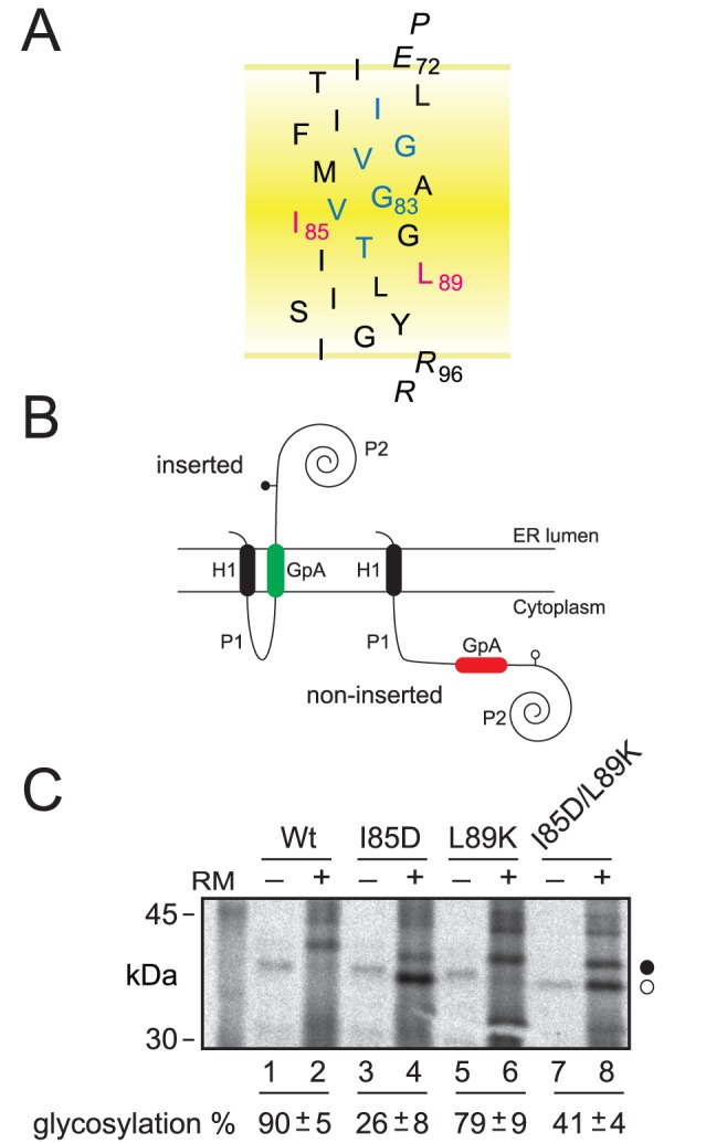
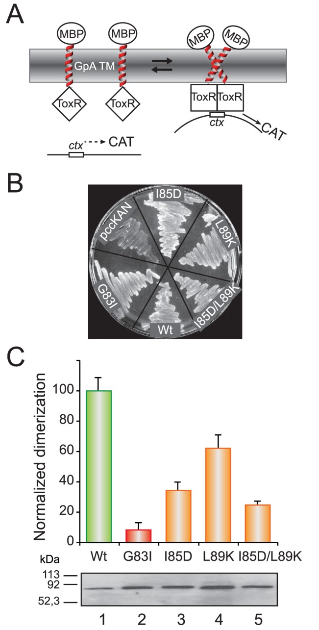
The vast majority of membrane proteins are anchored to biological membranes through hydrophobic α-helices. Sequence analysis of high-resolution membrane protein structures show that ionizable amino acid residues are present in transmembrane (TM) helices, often with a functional and/or structural role. Here, using as scaffold the hydrophobic TM domain of the model membrane protein glycophorin A (GpA), we address the consequences of replacing specific residues by ionizable amino acids on TM helix insertion and packing, both in detergent micelles and in biological membranes. Our findings demonstrate that ionizable residues are stably inserted in hydrophobic environments, and tolerated in the dimerization process when oriented toward the lipid face, emphasizing the complexity of protein-lipid interactions in biological membranes.


Conflict of interest statement

Competing Interests: The authors have declared that no competing interests exist.

Figures

Figure 1
Figure 1. Venn diagram of TM segments (the central 19 residues) containing charged residues: Asp (D, red), Lys (K, blue), Glu (E, yellow) and Arg (R, green).
The value in parenthesis is the total TM helices that contain at least one of such residues. The values inside the ellipses indicate the number of TM helices in each combination of these four amino acids. For example, there are 57 TM helices with only Lys as a charged residue, 12 helices with only Lys and Asp, 7 helices with Lys, Asp and Glu, and 3 helices with all four ionizable residues.
Figure 2
Figure 2. Structure of the calcium ATPase 1.
Left panel: cartoon representation of sarcoplasmic/endoplasmic reticulum calcium ATPase 1 (PDB ID: 1SU4) with cytosolic domain in the up side and transmembrane aspartic 59 and arginine 63 residues in spheres representation (C atom gray, O atom red and N atom blue). Membrane boundaries (dashed red lines) were obtained from the PPM Server . Right panel: zoom view centered on the salt bridge between Asp59 and Arg63, dashed pink lines indicate O to N atom distances.
Figure 3
Figure 3. Dimerization in SDS micelles.
(A) Helical wheel projection of GpA TM sequence. The residues associated with dimer formation as defined by Orzaez et al. are shown in blue. Non-interacting residues susceptible of ionizable residue substitution are shown in magenta. (B) Green colored bars denoted dimerization levels similar to wild-type sequence. Bars for intermediate dimerization levels (≈40%) are colored orange. Red colored bars denote non-dimerizing sequences (dimerization<3%). (C) SDS-PAGE analysis of GpA mutants. Chimeric proteins were purified in the presence of SDS and analyzed by PAGE. Positions of the monomer and dimer of the chimeras are marked on the right as single and double helices, respectively.
Figure 4
Figure 4. Insertion of GpA-derived segments into microsomal membranes.
(A) Model of the GpA TM wild-type sequence. GpA residues involved in dimer formation are blue, the hydrophobic residues replaced to ionizable residues are magenta, and flanking residues are shown in italic. (B) Membrane topology of Lep chimeras. The second TM segment of Lep was replaced by the GpA TM amino acid sequence (gray). The glycosylation acceptor site located in the beginning of the P2 domain will be modified only if GpA-derived TM sequence inserts into the membrane. (C) In vitro translation in the presence of rough microsomal membranes (RM). Proper insertion of the GpA-derived TM sequences results in an increase in molecular mass of about 2.5 kDa relative to the observed molecular mass of the proteins expressed in the absence of microsomes. Bands of nonglycosylated and glycosylated proteins are indicated by white and black dots, respectively. Average ± s.d. of glycosylation results from four independent experiments are shown at the bottom.
Figure 5
Figure 5. Dimerization in E. coli membranes.
(A) Schematic representation of the ToxCAT assay. ToxR domains (squares) can activate transcription of the reporter gene (CAT) if brought together by the GpA-derived TM domains (right). The maltose binding protein domain (ellipses) helps direct the insertion of the construct into the membrane, complements the malE mutation in the host cells, and serves as an epitope for quantifying the expression level of fusion protein. (B) Complementation assays for wild-type and selected mutant ToxR(GpA)MBP fusion constructs. NT326 cells (malE deficient) carrying various constructs were streaked on a plate with maltose as the sole carbon source and grown for three days at 37°C. All ToxR(GpA)MBP chimeras permit growth of NT326 cells on maltose, while control transformants (pccKAN) do not. (C) Normalized dimerization of the indicated TM domain variants as measured by CAT-ELISA relative to the wild-type GpA TM domain. Bars for intermediate dimerization and non-dimerizing levels are colored orange and red, respectively. Average ± s.d. of results from four independent experiments are shown. Levels of expression of selected ToxR(GpA)MBP constructs as analyzed by immunoblotting are shown at the bottom.

References

    1. Popot JL, Engelman DM (1990) Membrane protein folding and oligomerization - The 2-stage model. Biochemistry 29: 4031–4037. - PubMed
    1. Hessa T, Kim H, Bihlmaier K, Lundin C, Boekel J, et al. (2005) Recognition of transmembrane helices by the endoplasmic reticulum translocon. Nature 433: 377–381. - PubMed
    1. Baeza-Delgado C, Marti-Renom MA, Mingarro I (accepted) Structure-based statistical analysis of transmembrane segments. Eur Biophys J DOI 10.1007/s00249-012-0813-9. - PubMed
    1. Martinez-Gil L, Perez-Gil J, Mingarro I (2008) The surfactant peptide KL4 sequence is inserted with a transmembrane orientation into the endoplasmic reticulum membrane. Biophys J 95: L36–38. - PMC - PubMed
    1. Hessa T, Meindl-Beinker NM, Bernsel A, Kim H, Sato Y, et al. (2007) Molecular code for transmembrane-helix recognition by the Sec61 translocon. Nature 450: 1026–1030. - PubMed

Publication types

MeSH terms